. 2007 May;8(5):486-99.
doi: 10.1111/j.1600-0854.2007.00548.x.

The ternary Rab27a-Myrip-Myosin VIIa complex regulates melanosome motility in the retinal pigment epithelium

Affiliations

The ternary Rab27a-Myrip-Myosin VIIa complex regulates melanosome motility in the retinal pigment epithelium

Vanda S Lopes et al. Traffic. 2007 May.

Abstract

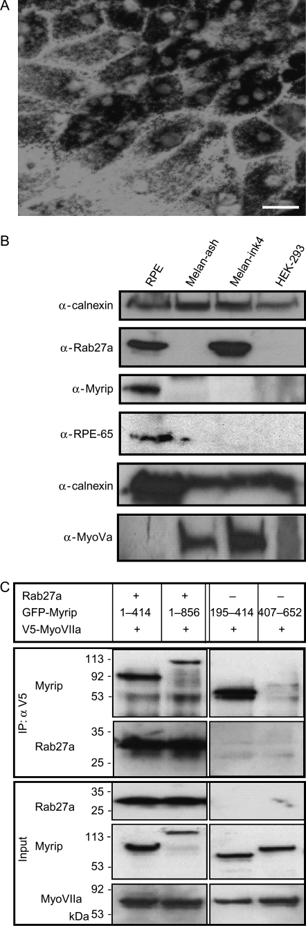
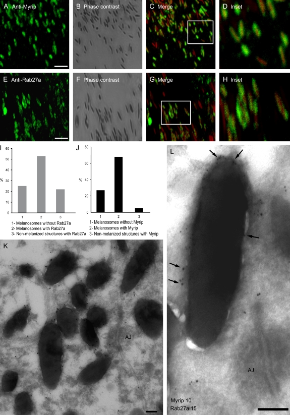
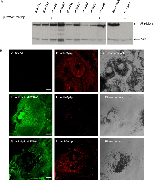
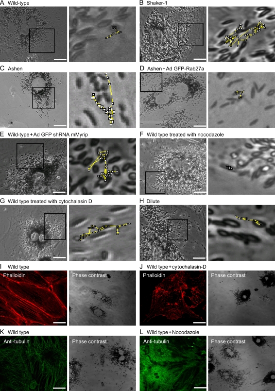
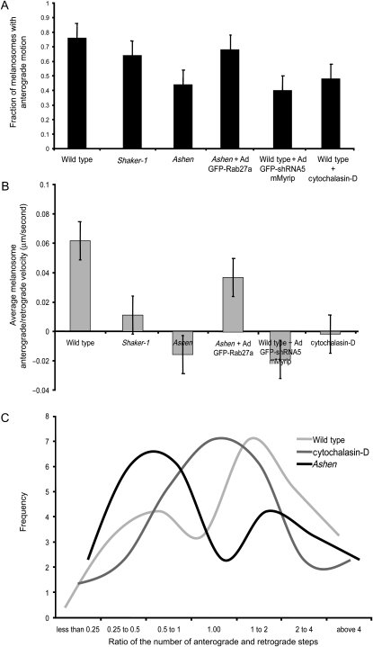
The retinal pigment epithelium (RPE) contains melanosomes similar to those found in the skin melanocytes, which undergo dramatic light-dependent movements in fish and amphibians. In mammals, those movements are more subtle and appear to be regulated by the Rab27a GTPase and the unconventional myosin, Myosin VIIa (MyoVIIa). Here we address the hypothesis that a recently identified Rab27a- and MyoVIIa-interacting protein, Myrip, promotes the formation of a functional tripartite complex. In heterologous cultured cells, all three proteins co-immunoprecipitated following overexpression. Rab27a and Myrip localize to the peripheral membrane of RPE melanosomes as observed by immunofluorescence and immunoelectron microscopy. Melanosome dynamics were studied using live-cell imaging of mouse RPE primary cultures. Wild-type RPE melanosomes exhibited either stationary or slow movement interrupted by bursts of fast movement, with a peripheral directionality trend. Nocodazole treatment led to melanosome paralysis, suggesting that movement requires microtubule motors. Significant and similar alterations in melanosome dynamics were observed when any one of the three components of the complex was missing, as studied in ashen- (Rab27a defective) and shaker-1 (MyoVIIa mutant)-derived RPE cells, and in wild-type RPE cells transduced with adenovirus carrying specific sequences to knockdown Myrip expression. We observed a significant increase in the number of motile melanosomes, exhibiting more frequent and prolonged bursts of fast movement, and inversion of directionality. Similar alterations were observed upon cytochalasin D treatment, suggesting that the Rab27a-Myrip-MyoVIIa complex regulates tethering of melanosomes onto actin filaments, a process that ensures melanosome movement towards the cell periphery.


Figures

Figure 1
Figure 1. Formation of a Rab27a–Myrip–MyoVIIa tripartite complex
A) Phase contrast image of cultured primary wild-type RPE cells at low magnification (× 40); Bar = 20 μm. B) Protein extracts prepared from wild-type murine RPE primary cells, ashen melanocytes, melan-ink (wild type) melanocytes and HEK-293 were separated by SDS–PAGE and immunoblotted for Rab27a, Myrip, RPE65, MyoVa and calnexin (as a loading control) as described under Materials and Methods. C) Plasmids encoding V5-MyoVIIa tail, GFP-Myrip and Rab27a were co-transfected into COS-7 cells and the expressed proteins were immunoprecipitated with anti-V5 or anti-Rab27a antibody as described under Materials and Methods. The bottom panels show total lysates after transfection, indicating transfection efficiencies. Top panels show immunoprecipitated proteins revealed by the indicated antibodies.
Figure 2
Figure 2. Subcellular localization of Rab27a and Myrip in RPE
Wild-type murine RPE cells were fixed, permeabilized and labelled with antibodies to Myrip (A) or Rab27a (E), as indicated. B and F) These panels show the corresponding phase contrast images and (C) and (G) are merged fluorescent and phase contrast images. D and H) These panels show boxed regions at higher magnification. Bar = 4 μm. Quantification of melanized and non-melanized structures staining with anti-Rab27a (I) and anti-Myrip (J) are represented. K and L) These panels show immunoelectron microscopy sections of wild-type murine RPE from retinal sections labelled with anti-Myrip antibody and 15 nm protein A–gold (G) or anti-Myrip antibody (10 nm gold) and anti-Rab27a (15 nm gold indicated by arrows) (H). AJ, adherens junction. Bar = 200 nm.
Figure 3
Figure 3. Efficiency of shRNAs to KD Myrip expression
A) HEK-293FT cells were co-transfected with pENTR/CMV-V5-Myrip and pENTR/U6-shRNA. Protein extracts were subjected to immunoblot using anti-V5 antibody. B) Wild-type RPE cells were fixed, permeabilized and subjected to immunofluorescence using anti-Myrip antibody. Wild-type RPE cells expressing no GFP-shRNA (panels A–C), GFP-Myrip shRNA5 (panels D–F) and GFP non-specific shRNA4 (panels G–H) were labelled with Myrip antibody, as indicated. Bar = 20 μm.
Figure 4
Figure 4. Live-cell imaging of RPE primary cultures and distribution of MTs and actin filaments
Phase contrast images of primary RPE cells at high magnification (× 100) are shown. Zoomed images showing representative trajectories drawn during the entire time frame collected during live-cell imaging illustrates the movement of melanosomes in representative wild-type RPE cell (A), shaker-1 RPE cell (B), ashen RPE cell (C), ashen RPE cell transduced with Ad GFP-Rab27a (D), wild-type RPE cell transduced with Ad shRNA5 Myrip (E), wild-type RPE cell treated with nocodazole (F) or cytochalasin D (G) and dilute RPE cell (H). Regions used for the movies are marked for each case. Wild-type murine RPE cells were fixed, permeabilized and labelled with phalloidin (I) or anti-tubulin (K), as indicated. Cells treated with 10 μm cytochalasin D were also stained with phalloidin (J), while cells incubated with 10 μm nocodazole were labelled with anti-tubulin (L). Bar = 20 μm.
Figure 5
Figure 5. Analysis of melanosome dynamics in RPE primary cultures
Representative kymograph tracings illustrating movement of individual melanosomes are shown for the indicated RPE cultures. Arrows indicate bursts of rapid movement. Tracing was started around 10–15 seconds before rapid movement was noticed.
Figure 6
Figure 6. Melanosome motion directionality
Histograms representing the fraction of melanosomes with centrifugal movement (A) and average melanosome anterograde/retrograde velocity (μm/second) (B) are shown for the indicated RPE cultures (mean ± standard deviation). C) Frequency of the ratio of number centrifugal/centripetal steps performed in a population of 25 melanosomes during a 30-second period.

References

    1. Futter CE. The molecular regulation of organelle transport in mammalian retinal pigment epithelial cells. Pigment Cell Res. 2006;19:104–111. - PubMed
    1. Strauss O. The retinal pigment epithelium in visual function. Physiol Rev. 2005;85:845–881. - PubMed
    1. Jeffery G. The albino retina: an abnormality that provides insight into normal retinal development. Trends Neurosci. 1997;20:165–169. - PubMed
    1. Futter CE. The role of Rab27a in the regulation of melanosome distribution within retinal pigment epithelial cells. Mol Biol Cell. 2004;15:2264–2275. - PMC - PubMed
    1. Gibbs D, et al. Role of myosin VIIa and Rab27a in the motility and localization of RPE melanosomes. J Cell Sci. 2004;117:6473–6483. - PMC - PubMed

Publication types

MeSH terms