. 2007 Jun 8;357(3):792-9.
doi: 10.1016/j.bbrc.2007.04.022. Epub 2007 Apr 13.

EHD1 regulates cholesterol homeostasis and lipid droplet storage

Affiliations

EHD1 regulates cholesterol homeostasis and lipid droplet storage

Naava Naslavsky et al. Biochem Biophys Res Commun. .

Abstract

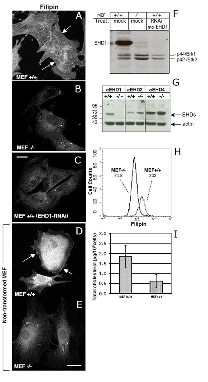
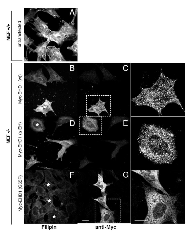
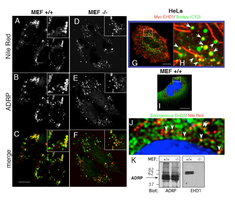
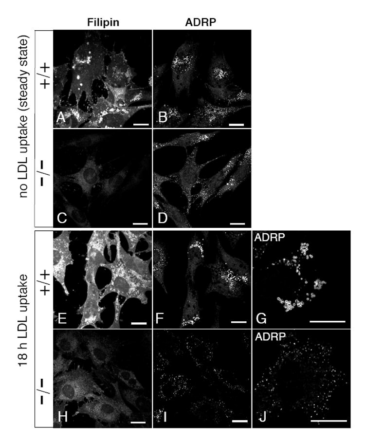
Endocytic transport is critical for the subcellular distribution of free cholesterol and the endocytic recycling compartment (ERC) is an important organelle that stores cholesterol and regulates its trafficking. The C-terminal EHD protein, EHD1, controls receptor recycling through the ERC and affects free cholesterol distribution in the cell. We utilized embryonic fibroblasts from EHD1 knockout mice (Ehd1(-/-)MEF) and SiRNA in normal MEF cells to assess the role of EHD1 in intracellular transport of cholesterol. Surprisingly, Ehd1(-/-)MEFs displayed reduced levels of esterified and free cholesterol, which returned to normal level upon re-introduction of wild-type, but not dysfunctional EHD1. Moreover, triglyceride and cholesterol storage organelles known as 'lipid droplets' were smaller in size in cells lacking EHD1, indicating that less esterified cholesterol and triglycerides were being stored. Decreased cellular cholesterol and reduced lipid droplet size in Ehd1(-/-)MEFs correlated with ineffectual cholesterol uptake via LDL receptor, suggesting involvement of EHD1 in LDL receptor internalization.


Figures

Fig. 1
Fig. 1. Decreased free cellular cholesterol in MEF cells lacking EHD1
(A-C) SV40-transformed MEF cells, and (D and E) primary, non-transformed MEF cells were plated on glass cover–slips for 24 h. Ehd1+/+ MEF cells (C) were treated with EHD1-RNAi for 48 h, whereas Ehd1+/+ and Ehd1-/- MEF cells were mock-treated (A-B, respectively). Cells were fixed and free cholesterol was visualized by Filipin staining (A-E). Arrows depict plasma membrane cholesterol. (F) Lysates from mock-treated Ehd1+/+ and Ehd1-/-MEF cells, and Ehd1+/+ MEF cells treated with EHD1-RNAi (as in C) were lysed and separated by SDS-PAGE and immunoblotted with rabbit anti-EHD1 and with anti-Erk 1/2 to show levels of protein loading. (G) The protein levels of other C-terminal EHD paralogs were analyzed in Ehd1+/+ and Ehd1-/-MEF cells. Lysates with equivalent protein concentrations were separated by SDS PAGE and immunoblotted with antibodies for EHD1, EHD2, EHD4, and with anti-actin as a control. (H) Flow cytometry analysis of free cholesterol was performed on at least 10,000 Ehd1+/+ and Ehd1-/- MEF cells stained with Filipin in the presence of saponin to permeabilize the cells. Mean fluorescence of each cell population is depicted in the graph. (I) Total cellular cholesterol levels (cholesterol and cholesteryl-ester) of Ehd1+/+ and Ehd1-/- MEF cells were measured by fluorometric assay. The mean and standard deviation are from 3 independent experiments, each utilizing 1 × 10⁶ cells. Bar, 10 μm.
Fig. 2
Fig. 2. Introduction of wt EHD1, but not mutant EHD1, into MEF -/- cells rescues cellular free cholesterol levels
Ehd1+/+ MEF cells (A) and Ehd1-/- MEF cells (B-G) were plated on glass cover-slips for 18 h. Ehd1-/- MEF cells were then transfected with wild-type Myc-EHD1 (B, C), Myc-EHD1 ΔEH (D, E), and myc-EHD1 G65R (F-G) for 48 h in the presence of 5 mM butyric acid for the last 18 h of transfection to enhance expression of the transfected proteins. After fixation and permeabilization, cells were stained with Filipin. Myc-EHD1 transfected proteins were identified with anti-Myc antibodies, followed by 568-Alexa goat anti-mouse IgG. Transfected cells stained with anti-Myc are enlarged to illustrate a more detailed staining pattern (outlined by ‘dashed rectangles’; insets for C, E and G). Stars depict transfected cells. Bar 10 μm.
Fig. 3
Fig. 3. Lack of EHD1 alters size and distribution of lipid droplets in Ehd1-/- MEF cells, and EHD1 and lipids droplets partially co-localize in Ehd1+/+MEF cells
(A-C) Ehd1+/+ MEFs and (D-F) Ehd1-/- MEFs were plated on glass cover-slips for 24 h in fatty-acid free medium containing 100 μM oleic acid. After fixation and permeabilization, lipid droplets were co-stained with Nile Red and anti-ADRP, followed by Cy2-Donkey anti-Guinea pig IgG. Insets depict the size of a cluster of lipid droplets. (G and inset; H) Myc-EHD1-transfected HeLa cells were grown on glass cover-slips in fatty-acid free medium containing 4 μg/ml Bodipy-(C12)-Fatty acid (visualized in green) for 18 h at 37°C. After fixation, the cells were incubated with anti-Myc followed by 568-goat anti-mouse IgG to detect Myc-EHD1 (red). (I and inset; J) Untransfected Ehd1+/+ MEF cells were incubated with fatty-acid free medium supplemented with 100 μM oleic acid for 18 h at 37°C, and fixed. The cells were co-stained (in the presence of saponin) with Nile Red to visualize lipid droplets, DAPI to mark the nuclei (blue), and with rabbit anti-EHD1 followed by 488-Alexa anti-rabbit IgG to detect endogenous EHD1 (green). Arrows mark EHD1 localized to Nile Red labeled lipid droplets, and arrowheads depict alignment of lipid droplets with EHD1-containing tubular membranes. (K) Equal numbers of Ehd1+/+ and Ehd1-/-MEFs were lysed, separated on SDS-PAGE and immunoblotted with either anti-ADRP (left panel), or anti-EHD1 (right panel). Bars, 10 μm.
Fig. 4
Fig. 4. Reduced delivery of LDL-derived cholesterol to lipid droplets in cells lacking EHD1
Ehd1+/+ MEF cells (A, B, E-G) and Ehd1-/-MEF cells (C, D, H-J) were plated on glass cover-slips for 18 h in complete media. For LDL uptake (E-J) the complete media was replaced by media containing fatty-acid free serum, and supplemented with 30 μg/ml serum-derived LDL for 18 h at 37°C prior to fixation. As a control (A-D) MEF cells growing in complete medium (without LDL) were fixed. All cover slips were permeabilized and stained with Filipin and anti-ADRP, followed by 568-conjugated goat-anti-Guinea pig IgG. Insets (G) and (J) depict ADRP stain of lipid droplets in a single-cell shown at higher magnification. Bars, 10 μm.

References

    1. Brown MS, Goldstein JL. A receptor-mediated pathway for cholesterol homeostasis. Science. 1986;232:34–47. - PubMed
    1. Chang TY, Chang CC, Cheng D. Acyl-coenzyme A:cholesterol acyltransferase. Annu Rev Biochem. 1997;66:613–638. - PubMed
    1. Maxfield FR, Wustner D. Intracellular cholesterol transport. J Clin Invest. 2002;110:891–898. - PMC - PubMed
    1. Soccio RE, Breslow JL. Intracellular cholesterol transport. Arterioscler Thromb Vasc Biol. 2004;24:1150–1160. - PubMed
    1. Londos C, Brasaemle DL, Schultz CJ, Segrest JP, Kimmel AR. Perilipins, ADRP, and other proteins that associate with intracellular neutral lipid droplets in animal cells. Semin Cell Dev Biol. 1999;10:51–58. - PubMed

MeSH terms