. 2007 Jun 8;369(3):794-811.
doi: 10.1016/j.jmb.2007.03.061. Epub 2007 Mar 30.

Crystal structure and allosteric regulation of the cytoplasmic Escherichia coli L-asparaginase I

Affiliations

Crystal structure and allosteric regulation of the cytoplasmic Escherichia coli L-asparaginase I

Mi-Kyung Yun et al. J Mol Biol. .

Abstract

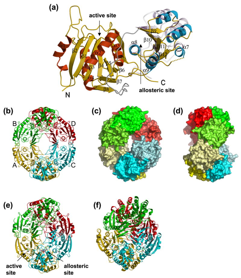
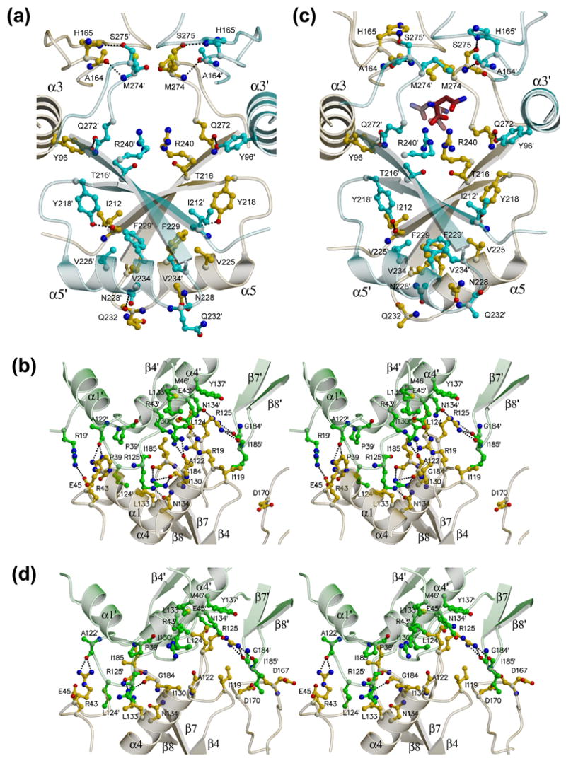
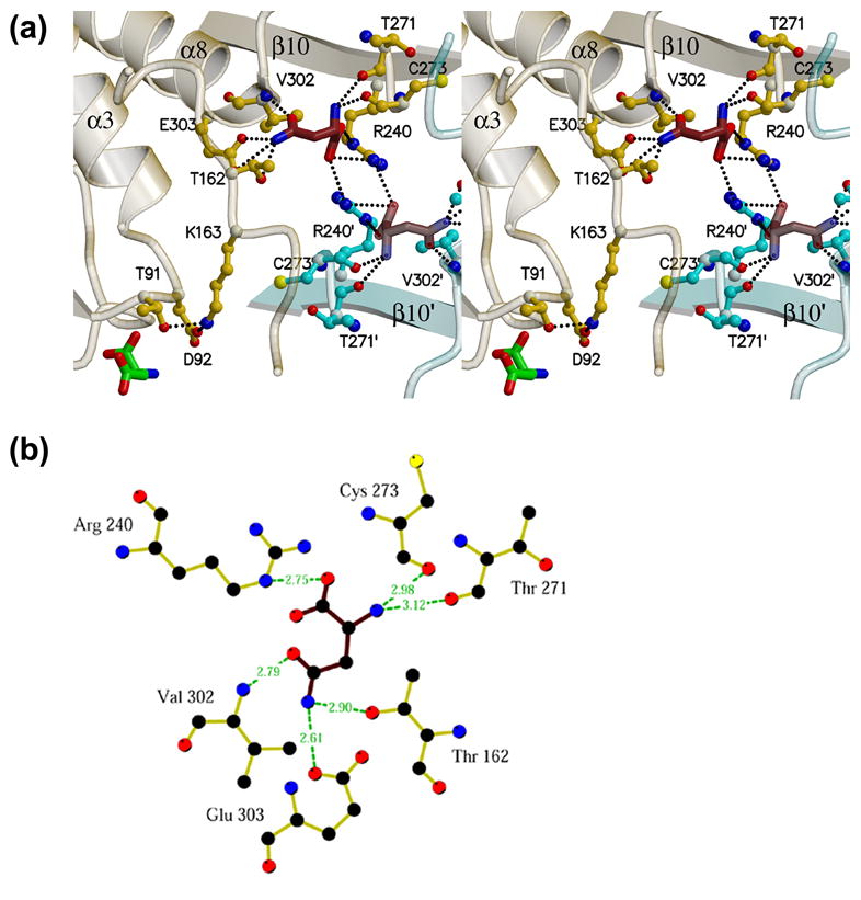
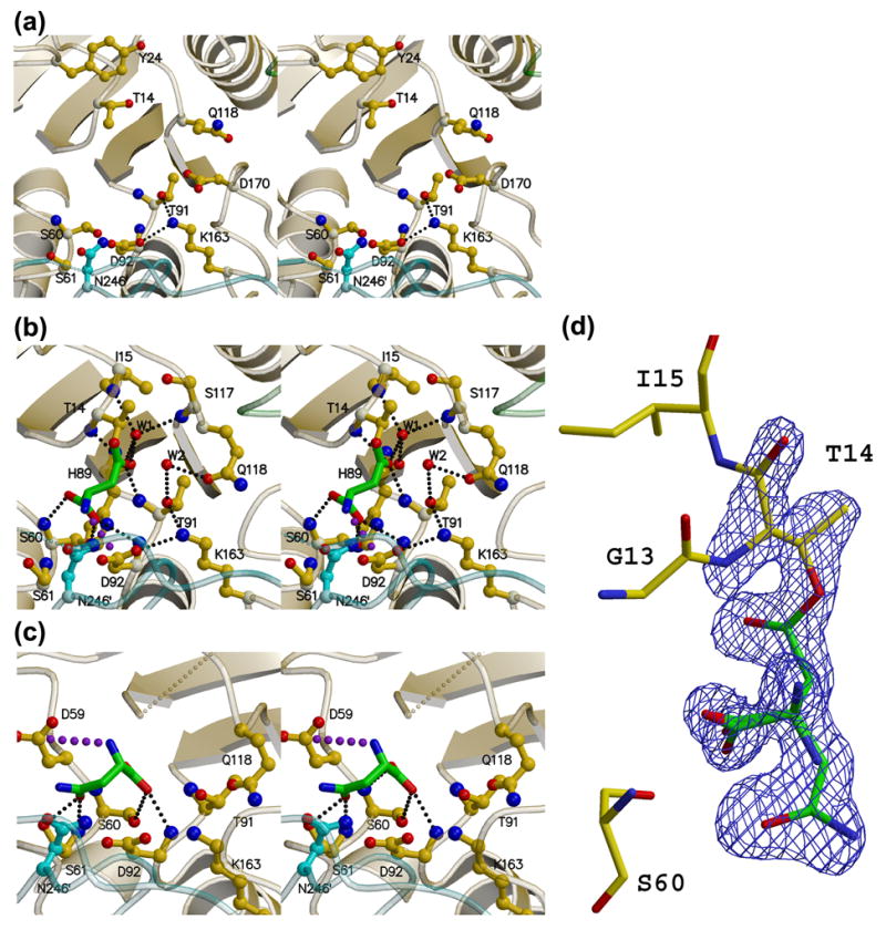
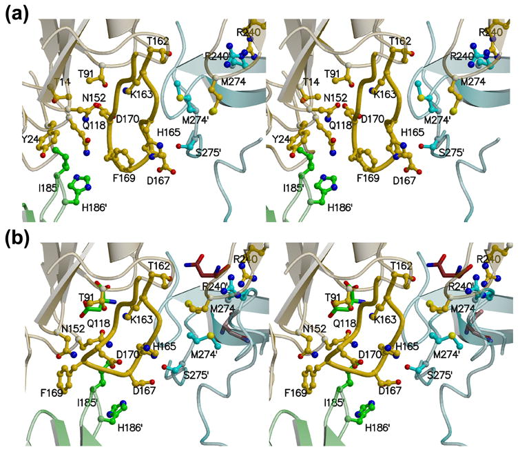
AnsA is the cytoplasmic asparaginase from Escherichia coli involved in intracellular asparagine utilization. Analytical ultracentifugation and X-ray crystallography reveal that AnsA forms a tetrameric structure as a dimer of two intimate dimers. Kinetic analysis of the enzyme reveals that AnsA is positively cooperative, displaying a sigmoidal substrate dependence curve with an [S](0.5) of 1 mM L-asparagine and a Hill coefficient (n(H)) of 2.6. Binding of L-asparagine to an allosteric site was observed in the crystal structure concomitant with a reorganization of the quarternary structure, relative to the apo enzyme. The carboxyl group of the bound asparagine makes salt bridges and hydrogen bonds to Arg240, while the N(delta2) nitrogen interacts with Thr162. Mutation of Arg240 to Ala increases the [S](0.5) value to 5.9 mM, presumably by reducing the affinity of the site for L-asparagine, although the enzyme retains cooperativity. Mutation of Thr162 to Ala results in an active enzyme with no cooperativity. Transmission of the signal from the allosteric site to the active site appears to involve subtle interactions at the dimer-dimer interface and relocation of Gln118 into the vicinity of the active site to position the probable catalytic water molecule. These data define the structural basis for the cooperative regulation of the intracellular asparaginase that is required for proper functioning within the cell.


Figures

Figure 1
Figure 1. Sequence alignment of selected asparaginases
The alignment and secondary structure locations are with respect to the E. coli type I (AnsA) enzyme described in this paper. The linker region that joins the N- and C-terminal domains of the structure is indicated. Regions of sequence identity and sequence similarity are shown in blue boxes and blue text, respectively. The conserved tyrosine with a putative catalytic role is indicated by black arrowheads (residue 24 in ecAnsA). The sequences of three intracellular AnsA enzymes and three extracellular AnsB enzymes are shown; ec, E. coli; ph, Pyrococcus horikoshii; sc, Saccharomyces cerevisiae; er, Erwinia chrysanthemi. For clarity, the signal sequences at the N-termini of AnsB enzymes are not shown, and an extended N-terminal region (amino acids 1–46) in scAnsA has been removed. The figure was generated with ESPript 2.2.
Figure 2
Figure 2. Overall structure of E. coli AnsA
(a) A ribbon representation of the apo AnsA monomer showing the secondary structure elements. The N-terminal domain α-helices are shown in red, the C-terminal domain α-helices are blue and the β-strands are in yellow in both domains of the molecule. The linker region is shown in grey. Shown semi-transparently is the position of the C-terminal domain relative to the fixed N-terminal domain in the structure of AnsA complexed with L-asparagine. Note that the movement is a rotation around helix α8 at the domain interface. (b–d) The tetrameric structure of apo AnsA with monomers A, B, C and D (labeled in b) in yellow, green, cyan and red, respectively. (b) A ribbon representation; (c) a surface representation showing the donut shape with the hole in the center; (d) a 90° rotation of (c) showing the tight (top) and loose (front, middle) interfaces. In (b) and (c), the N- and C-terminal domains are shaded differently for clarity. (e) The tetrameric structure of AnsA complexed with L-asparagine in a ribbon representation showing aspartate (green) at the four active sites and asparagine (brown) at the four allosteric sites (one of each site is indicated). Compared to the apo structure (b), the tetramer is more compact and the central hole is smaller. (f) The tetrameric structure of E. coli AnsB in ribbon representation in which the central hole is absent due to a more orthogonal packing of tight dimers.
Figure 3
Figure 3. Monomer-monomer interfaces in the AnsA tetramer
Panels (a) and (c) show the intimate dimer (A/C) interface in the apo structure and the asparagine-bound structure, respectively. Panels (b) and (d) show stereoviews of the loose dimer (A/B) interface in the apo structure and the asparagine-bound structure, respectively. Monomer A is yellow, B is green and C is in cyan, and the two allosteric asparagine molecules in (c) have red carbon atoms. Details are provided in the text.
Figure 4
Figure 4. The AnsA allosteric asparagine binding pocket
(a) The pocket is at the tight dimer interface (between monomers A and C in this view) at the N-terminus of helix α8 and spanning dyad-related arginines 240 and 240′. Details of the interaction are provided in the text. Elements of monomer A and C are shown in yellow and cyan, respectively. Note that two dyad-related pockets are visible in this view, as well as the active site aspartate in monomer A adjacent to Thr91. Note also that Thr162 and Lys163 connect the allosteric and active sites within one monomer. (b) Diagrammatic representation of the hydrogen bonding interactions of asparagine (brown bonds) in the allosteric site (yellow bonds). Figure was generated with Ligplot.
Figure 5
Figure 5. Analytical Ultracentrifugation of AnsA
(a) Absorbance scans at 280 nm at equilibrium are plotted versus the distance from the axis of rotation for AnsA. Protein samples were centrifuged at 4 °C for at least 48 h at 5,000 (red circle), 7,000 (green triangle), and 10,000 (black square) rpm. The solid lines represent the global nonlinear least squares best-fit of all the data to a single molecular species. For clarity only the sample with a loading protein concentration of 0.5 mg/ml is shown. Residuals of the fit at all rotor speeds are also shown and the r.m.s. deviation is 0.005 absorbance units. (b) The s-values of the c(s) distribution were converted to standard condition s20,w-values (20 °C, water as solvent), and plotted as shown. The r.m.s. deviation is 0.010 fringes. Experiments were conducted at a loading protein concentration of 1.1 mg/ml at 4 °C and at a rotor speed of 50,000 rpm. The s20,w-value of AnsA is 7.60 S.
Figure 6
Figure 6. Stereoviews of the AnsA active site
(a) The apo enzyme (APOM). (b) The AnsA-asparagine complex showing the covalently attached product aspartate (green) at the active site. Water molecule W2 hydrogen bonded to Thr91 and Gln118 is ideally positioned to act as the nucleophile that will release the product (green). (c) The AnsA-asparagine complex showing asparagine (green) bound in the non-productive alternate conformation. In each panel, monomer A is yellow, B is green and C is cyan. Details of these interactions are provided in the text, but the key active site residues are Thr14, Thr91, Lys163 and Asp92. Note how Gln118 changes conformation as the tetramer is compacted in (b) versus (a). Note also the pseudo mirror symmetry in the active site that accommodates the two alternate binding modes in (b) and (c). Large purple dots indicate a salt bridge, while small, light gray dots indicate missing structural elements. (d) Electron density observed in the active site that was interpreted as overlapped aspartate and asparagine in the non-productive alternate conformation. The Fo-Fc simulated annealing omit map is displayed at a contour level of 4σ (blue).
Figure 7
Figure 7. Relative positioning of Gln118 and Asp170 at the AnsA active site
During the allosteric switch upon binding asparagine, the two loops containing Gln118 and Asp170 adjacent to the active site change conformation. As a result, Gln118 occupies the space adjacent to Thr91 and Lys163 that was previously occupied by Asp170 in the apo structure. (a) Stereoview of the apo active site. (b) Stereoview of the AnsA-asparagine active site. In each panel, monomer A is yellow, B is green and C is cyan. Note how the active site locale is at the convergence of three subunits. Details are provided in the text.

Similar articles

Cited by

References

    1. Cedar H, Schwartz JH. Localization of the two L-asparaginases in anaerobically grown Escherichia coli. J Biol Chem. 1967;242:3753–3755. - PubMed
    1. Schwartz JH, Reeves JY, Broome JD. Two L-asparaginases from E. coli and their action against tumors. Proc Natl Acad Sci U S A. 1966;56:1516–1519. - PMC - PubMed
    1. Spring KJ, Jerlstrom PG, Burns DM, Beacham IR. L-asparaginase genes in Escherichia coli: isolation of mutants and characterization of the ansA gene and its protein product. J Bacteriol. 1986;166:135–142. - PMC - PubMed
    1. Cedar H, Schwartz JH. Production of L-asparaginase II by Escherichia coli. J Bacteriol. 1968;96:2043–2048. - PMC - PubMed
    1. Casale TD, Sollitti P, Chesney RH. Cytoplasmic L-asparaginase: isolation of a defective strain and mapping of ansA. J Bacteriol. 1983;154:513–515. - PMC - PubMed

Publication types

LinkOut - more resources