. 2007 Aug 1;364(2):301-16.
doi: 10.1016/j.virol.2007.03.037. Epub 2007 Apr 23.

Silencing and complementation of reovirus core protein mu2: functional correlations with mu2-microtubule association and differences between virus- and plasmid-derived mu2

Affiliations

Silencing and complementation of reovirus core protein mu2: functional correlations with mu2-microtubule association and differences between virus- and plasmid-derived mu2

John Carvalho et al. Virology. .

Abstract

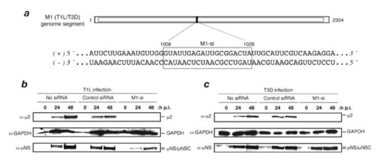
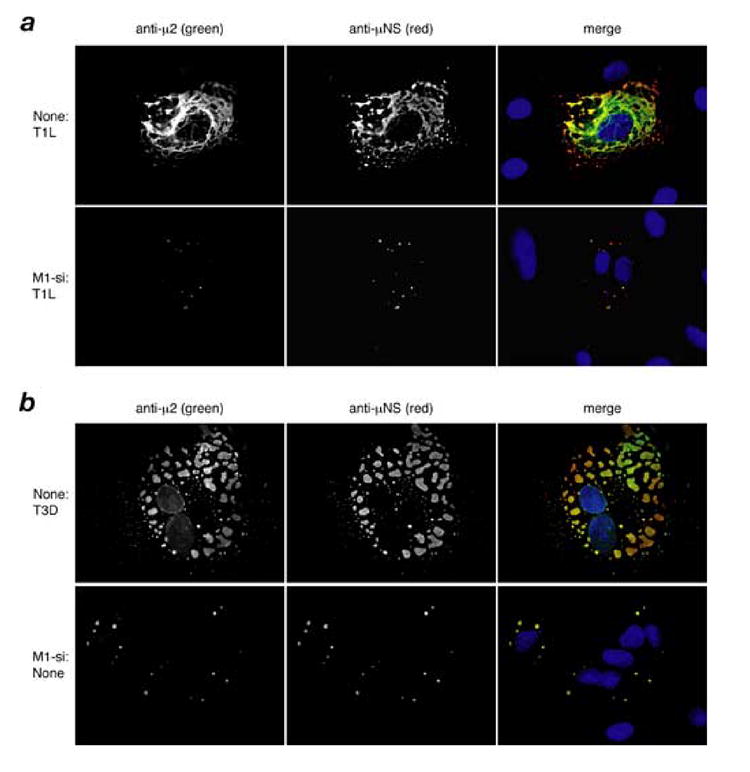
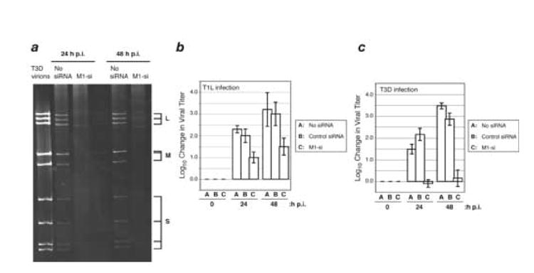
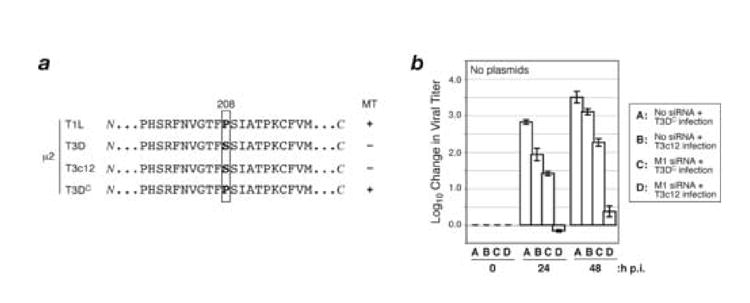
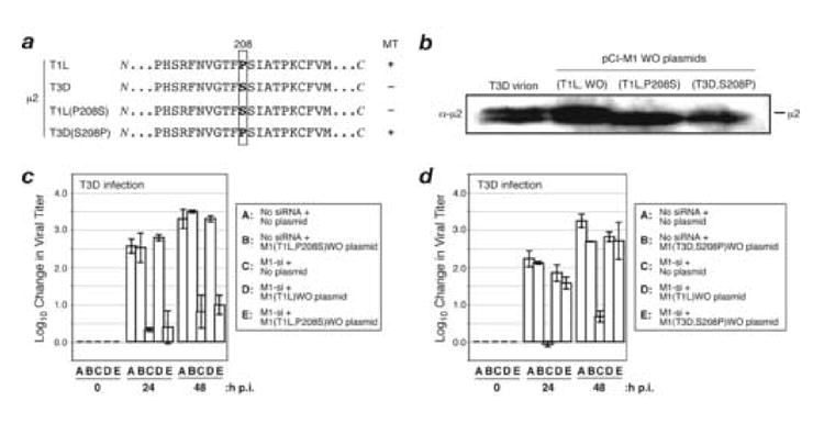
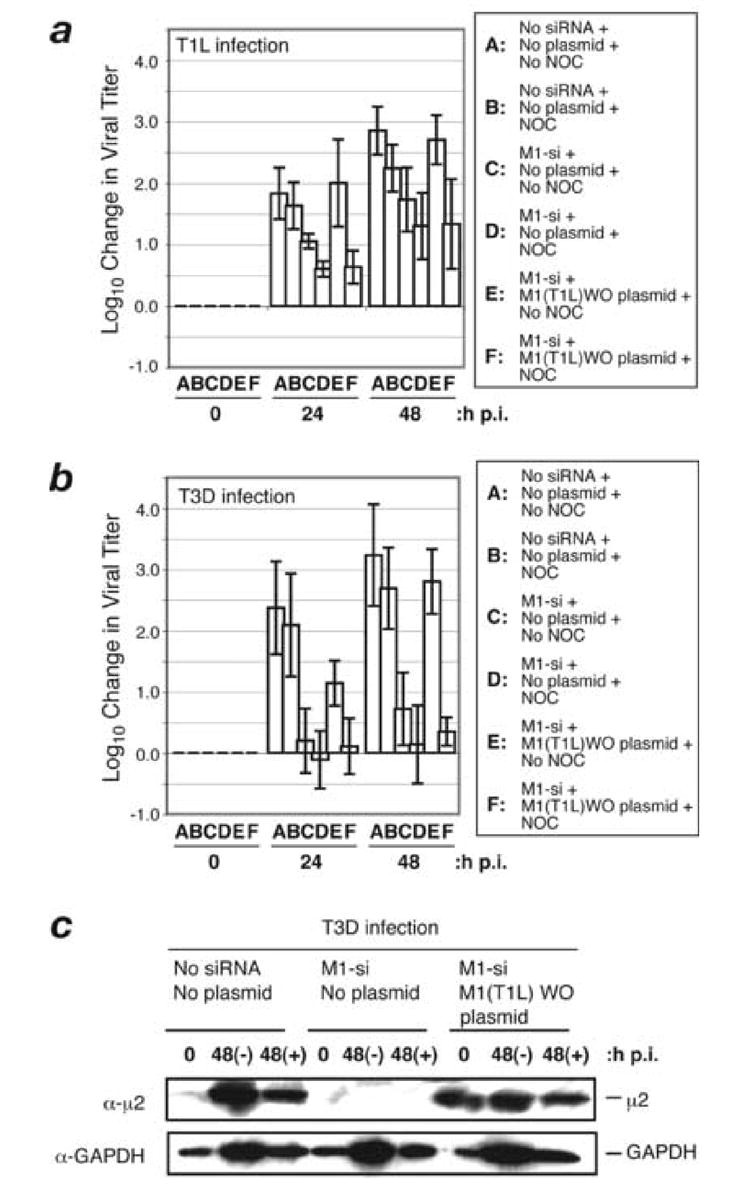
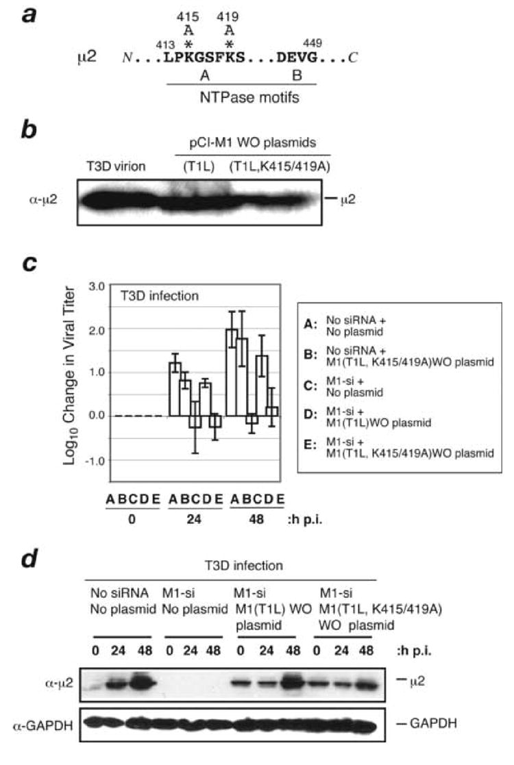
A low-copy component of mammalian reovirus particles is mu2, an 83-kDa protein encoded by the M1 viral genome segment and packaged within the viral core. Previous studies have identified mu2 as a nucleoside triphosphate phosphohydrolase (NTPase) as well as an RNA 5'-triphosphate phosphohydrolase (RTPase), putatively involved in reovirus RNA synthesis and/or 5'-capping. Other studies have identified mu2 as a microtubule-binding protein, which also associates with the viral factory matrix protein muNS and thereby anchors the factories to cellular microtubules during infections by most reovirus strains. To extend studies of mu2 functions during infection, we tested a small interfering RNA (siRNA) directed against the M1 plus-strand RNAs of reovirus strains Type 1 Lang (T1L) and Type 3 Dearing (T3D). The siRNA strongly suppressed mu2 expression by either strain and reduced infectious yields in a strain-dependent manner. This first strain difference was genetically mapped to the M1 genome segment and tentatively assigned to a single mu2 sequence polymorphism, Pro/Ser208, which also determines a T1L-T3D strain difference in microtubule association. The siRNA-based defect in mu2 expression was rescued by plasmids, containing silent mutations in the siRNA-targeted sequence, which encoded either T1L or T3D mu2, but the growth defect was rescued only by T1L mu2. This second strain difference was also mapped to Pro/Ser208, in that swapping this one residue between T1L and T3D mu2 reversed the rescue phenotypes. Thus, the T1L-T3D strain difference in mu2-microtubule association was correlated not only with the extent of reduction in infectious yields by the siRNA but also with the extent of rescue by plasmid-derived mu2. In addition, the rescue capacity of T1L mu2 was abrogated by nocodazole treatment, providing independent evidence for the importance of mu2-microtubule association in plasmid-based rescue. In two separate cases, the results revealed functional differences between virus- and plasmid-derived mu2. Ala substitutions within the NTP-binding motif of T1L mu2 also abrogated its rescue capacity, suggesting that the NTPase or RTPase activity of mu2 is additionally required for effective viral growth.


Figures

Fig. 1
Fig. 1. Effect of siRNA (M1-si) targeting a shared sequence in the M1 plus-strand RNAs of reovirus strains T1L and T3D on μ2 expression levels
(A) The M1 genome sequence of T1L and T3D is shown in the region targeted by M1-si. The M1-si-targeted region is boxed and encompasses nucleotides 1008 to 1026. (B, C) CV-1 cells were transfected with siRNA and at 24 h p.t. were then infected with T1L (B) or T3D (C) reovirus. The cells were incubated for an additional 0, 24, or 48 h p.i., and cell-associated proteins were prepared for SDS-PAGE and immunoblotting. Protein was detected using specific antibodies against μ2, GAPDH, or μNS.
Fig. 2
Fig. 2. Effect of M1-si on expression of μNS and expansion of viral factories for T1L and T3D reoviruses
(A, B) CV-1 cells were transfected with no siRNA or M1-si, and at 24 h p.t. were then infected with T1L (A) or T3D (B) reovirus at 10 PFU/cell. At 24 h p.i., the cells were fixed and the localization of μ2 and μNS proteins were examined by immunostaining with specific antibodies to μ2 (directly conjugated to Alexa 488, green channel) and μNS (followed by secondary antibody conjugated to Alexa 594, red channel). Nuclei were counterstained with DAPI, as seen in the merge images (right column).
Fig. 3
Fig. 3. Effect of M1-si on dsRNA synthesis and infectious viral yields
(A) CV-1 cells were transfected with no siRNA or M1-si, and at 24 h p.t. were then infected with T3D reovirus at 10 PFU/cell. Cells were incubated for 24 or 48 h p.i., and samples were prepared for gel electrophoresis to detect the viral dsRNA genome segments. L, genome segments encoding λ proteins; M, genome segments encoding μ proteins; S, genome segments encoding σ proteins. (B, C) CV-1 cells were transfected with no siRNA, nonspecific control siRNA, or M1-si, and at 24 h p.t. were then infected with T1L (B) or T3D (C) reovirus at 10 PFU/cell. Cells were harvested at 0, 24, or 48 h p.i., and viral titers were determined by plaque assays. Log10 changes in viral titer relative to time 0 are indicated. Each bar represents the average obtained from three independent experiments, and error bars indicate the standard deviations.
Fig. 4
Fig. 4. Effect of M1-si on infectious viral yields of T3DC and T3c12 reoviruses
(A) Protein sequences for μ2 from reovirus strains T1L, T3D, T3DC, and T3c12. T1L and T3DC reoviruses have a Pro at μ2 residue 208 and encode μ2 proteins that strongly associate with microtubules (+); T3D and T3c12 reoviruses have a Ser at this position and encode a μ2 protein that weakly associates with microtubules (−). (B) CV-1 cells were transfected with no siRNA or M1-si, and at 24 h p.t. were then infected with T3DC or T3c12 reovirus at 10 PFU/cell. Cells were harvested at 0, 24, or 48 h p.i., and viral titers were determined by plaque assays. Log10 changes in viral titer relative to time 0 are indicated. Each bar represents the average obtained from three independent experiments, and error bars indicate the standard deviations.
Fig 5
Fig 5. Effect of plasmid-based expression of T1L and T3D μ2 on M1-si-based reduction of infectious viral yields
(A) Sequence of rescue plasmid encoding T1L and T3D M1 in the region targeted by M1-si. Asterisks indicate the nucleotides that were changed to encode silent (wobble) mutations. (B) CV-1 cells were transfected with 5 μg of either pCI-M1(T1L) or pCI-M1(T3D) rescue plasmids. In each case, these plasmids either contained wobble mutations in the M1-si-targeted sequence (designated as WO plasmids) or did not (designated as WT plasmids). The cells were incubated for an additional 24 h p.i., and cell-associated proteins were prepared for SDS-PAGE and immunoblotting. Protein was detected using specific antibodies against μ2. (C, D, E, F) CV-1 cells were transfected with no siRNA, M1-si, or M1-si in combination with a particular rescue plasmid, and at 24 h p.t. were then infected with T1L (C, F) or T3D (D, E) reovirus at 10 PFU/cell. Cells were harvested at 0, 24, or 48 h p.i., and viral titers were determined by plaque assays. Log10 changes in viral titer relative to time 0 are indicated. Each bar represents the average obtained from three independent experiments, and error bars indicate the standard deviations. (G) CV-1 cells were transfected with no siRNA, M1-si, or M1-si in combination with either M1(T1L)WT or M1(T1L)WO plasmid, and at 24 h p.t. were then infected with T3D reovirus. The cells were incubated for an additional 0, 24, or 48 h p.i., and cell-associated proteins were prepared for SDS-PAGE and immunoblotting. Protein was detected using specific antibodies against μ2 or GAPDH.
Fig. 6
Fig. 6. Effect of μ2 amino acid Pro/Ser208 on complementation of M1-si-based reduction of infectious viral yields
(A) Protein sequences for μ2 from reovirus strains T1L or T3D, or plasmids encoding T1L(P208S) or T3D(S208P) μ2. Box indicates the position of amino acid 208. T1L reovirus and plasmid encoding T3D(S208P) μ2 have a Pro at this position and encode a μ2 protein capable of strongly associating with microtubules (+); T3D reovirus and plasmid encoding T1L(P208S) μ2 have a Ser substituted at this position and weakly associate with microtubules (−). (B) CV-1 cells were transfected with 5 μg of either M1(T1L,P208S)WO or M1(T3D,S208P)WO rescue plasmids. The cells were incubated for an additional 24 h p.i., and cell-associated proteins were prepared for SDS-PAGE and immunoblotting. Protein was detected using specific antibodies against protein μ2. (C, D) CV-1 cells were transfected with no siRNA, rescue plasmid alone, M1-si alone, or M1-si in combination with a particular rescue plasmid, and at 24 h p.t. were infected with T3D reovirus at 10 PFU/cell. Cells were harvested at 0, 24, or 48 h p.i., and viral titers were determined by plaque assays. Log10 changes in viral titer relative to time 0 are indicated. Each bar represents the average obtained from three independent experiments, and error bars indicate the standard deviations.
Fig. 7
Fig. 7. Effect of nocodazole on complementation of M1-si-based reduction of infectious viral yields
(A, B) CV-1 cells were transfected with no siRNA, M1-si, or M1-si in combination with M1(T1L)WO plasmid, and at 24 h p.t. were infected with T1L (A) or T3D (B) reovirus at 10 PFU/cell. At 6 h p.i., a subset of the infected cells were treated with 10 μM nocodazole and incubated further. Cells were harvested at 0, 24, or 48 h p.i., and viral titers were determined by plaque assays. Log10 changes in viral titer relative to time 0 are indicated. Each bar represents the average obtained from three independent experiments, and error bars indicate the standard deviations. (C) CV-1 cells were transfected with no siRNA, M1-si, or M1-si in combination with M1(T1L)WO plasmid, and at 24 h p.t. were infected with T3D reovirus at 10 PFU/cell. At 6 h p.i., a subset of the infected cells were treated with 10 μM nocodazole and incubated further. Samples were harvested at 0 and 48 h p.i., and cell-associated proteins were prepared for SDS-PAGE and immunoblotting. Protein was detected using specific antibodies against μ2 or GAPDH.
Fig. 8
Fig. 8. Effect of NTPase or RTPase function of μ2 protein on complementation of M1-si-based reduction of infectious viral yields
(A) Putative NTP-binding motifs in the T1L μ2 protein are shown. Asterisks indicate the two Ala-for-Lys substitutions in the mutant μ2 protein K415/419A. (B) CV-1 cells were transfected with 5 μg of M1(T1L) WO or M1(T1L,K415/419A)WO plasmid. Samples were harvested at 24 h p.i., and cell-associated protein was prepared for SDS-PAGE and immunoblotting. Protein was detected using specific antibodies against μ2. (C) CV-1 cells were transfected with no siRNA, M1(T1L,K415/419A)WO rescue plasmid, M1-si alone, M1-si plus M1(T1L)WO rescue plasmid, or M1-si plus M1(T1L,K415/419A)WO rescue plasmid, and at 24 h p.t. were infected with T3D reovirus at 10 PFU/cell. Cells were harvested at 0, 24, or 48 h p.i., and viral titers were determined by plaque assays. Log10 changes in viral titer relative to time 0 are indicated. Each bar represents the average obtained from three independent experiments, and error bars indicate the standard deviations. (D) CV-1 cells were transfected with no siRNA, M1-si, or M1-si in combination with M1(T1L)WO plasmid or M1(T1L,K415/419A)WO plasmid, and at 24 h p.t. were then infected with T3D reovirus. The cells were incubated for an additional 0, 24, or 48 h p.i., and cell-associated proteins were prepared for SDS-PAGE and immunoblotting. Protein was detected using specific antibodies against μ2 or GAPDH.

Similar articles

Cited by

References

    1. Becker MM, Goral MI, Hazelton PR, Baer GS, Rodgers SE, Brown EG, Coombs KM, Dermody TS. Reovirus σNS protein is required for nucleation of viral assembly complexes and formation of viral inclusions. J Virol. 2001;75:1459–1475. - PMC - PubMed
    1. Becker MM, Peters TR, Dermody TS. Reovirus σNS and µNS proteins form cytoplasmic inclusion structures in the absence of viral infection. J Virol. 2003;77:5948–5963. - PMC - PubMed
    1. Bodkin DK, Fields BN. Growth and survival of reovirus in intestinal tissue: role of the L2 and S1 genes. J Virol. 1989;63:1188–1193. - PMC - PubMed
    1. Brentano L, Noah DL, Brown EG, Sherry B. The reovirus protein μ2, encoded by the M1 gene, is an RNA-binding protein. J Virol. 1998;72:8354–8357. - PMC - PubMed
    1. Broering TJ, McCutcheon AM, Centonze VE, Nibert ML. Reovirus nonstructural protein μNS binds to reovirus cores, but does not inhibit their transcription activity. J Virol. 2000;74:5516–5524. - PMC - PubMed

Publication types

MeSH terms