

doi: 10.1016/j.jneuroim.2007.03.002.
Epub 2007 Apr 23.
Antigen specificity of clonally expanded and receptor edited cerebrospinal fluid B cells from patients with relapsing remitting MS
Affiliations
- PMID: 17451814
- PMCID: PMC2709235
- DOI: 10.1016/j.jneuroim.2007.03.002
Item in Clipboard
Antigen specificity of clonally expanded and receptor edited cerebrospinal fluid B cells from patients with relapsing remitting MS
J Neuroimmunol.
2007 May.
Abstract
We re-engineered the immunoglobulin rearrangements from clonally expanded CSF B cells of three Multiple Sclerosis patients as Fab fragments, and used three methods to test for their antigen (Ag) specificity. Nine out of ten Fab fragments were reactive to Myelin Basic Protein (MBP). The one Fab that did not react to MBP was a product of receptor editing. Two of the nine MBP reactive Fabs were also reactive to GFAP and CNPase, indicating that these clones were polyreactive. Targeting the mechanisms that allow these self-reactive B cells to reside in the CSF of MS patients may prove to be a potent immunotherapeutic strategy.
Figures
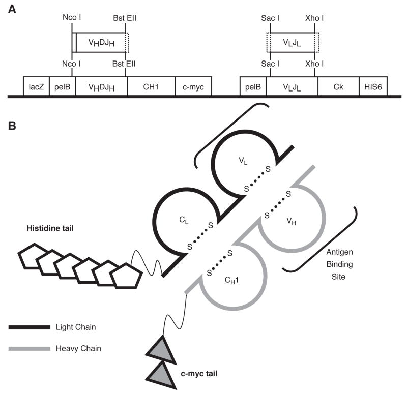
Panel A. Features of the D1.3v2 plasmid. The original FabD1.3mychis6 vector utilized the Pst I and Bst EII restriction digest enzymes to insert heavy chains into the heavy chain cassette. The Pst I site was converted to Nco I to prevent cutting within heavy chains, and is now called D1.3v2 Fab. Heavy and light chains are not linked by a disulfide bridge in this construct. Panel B. Illustration of a properly expressed Fab protein, including the features described.
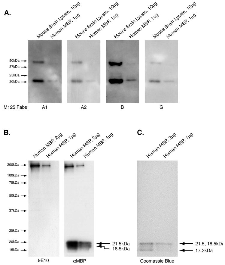
Panel A. Four sets of purified human MBP (1 ug/lane) and mouse Brain Lysate (10 ug/lane) were resolved by SDS-PAGE. Western blots were carried out as described in Materials and Methods using one of the four Fabs (M125-A1, -A2, -B or –G) as the primary Western blot antibody for one blot. All four of these Fabs are reactive to MBP. Panel B. Control blots showing reactivity of a commercially available anti-MBP antibody to human MBP (1 and 2 ug/lane), and the reactivity of the secondary 9E10, only. The bands at high molecular weight are due to incomplete reduction of the protein samples prior to electropheresis and correspond to the amount of protein loaded in the respective lanes. Panel C. Parallel SDS-PAGE of MBP (1 ug or 2 ug) resolved per lane then stained with coomassie blue to demonstrate the presence of MBP isoforms at 21.5 kDa, 18.5 kDa and 17.2 kDa.
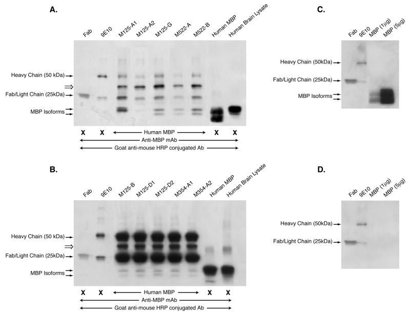
Panels A and B. Fabs were used as capture antibodies to MBP, and the resultant eluates of the IP reactions resolved on 4–20% PAGE gradient gels, transferred to PVDF membranes and probed with the commercially available anti-MBP antibody. Bands at 25 kDa and 50 kDa correspond to the light and heavy chains respectively of the c-myc antibody used to precipitate the Fabs bound to MBP. Fragments corresponding to MBP were detected as a doublet at 20 kDa with the exception of the M125-A1 IP, which seems to contain only one fragment. Note that the data represented in Panels A and B were not performed in the same experiment. Panel C. Components of the IP reaction (Fab, 9E10, and purified hMBP), which were resolved by SDS-PAGE, transferred to PVDF, and probed with anti-MBP followed by GAM-HRP. Panel D. Same filter was probed with GAM-HRP only. These experiments were performed 4 times with similar results.
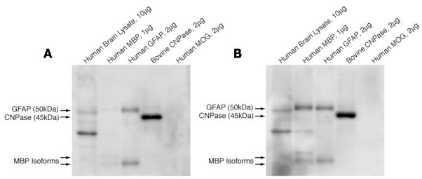
Purified antigens as indicated were resolved by SDS-PAGE, transferred to membranes, and WBs using either M125-D1 (Panel A) or –D2 Fabs (Panel B) as the primary probe were performed as described in materials and methods. Both clone members of M125-D recognize hCNPase and GFAP by WB. M125-D2 also recognizes MBP. Both Fabs also reacted to an unidentified 30 kDa protein in hBL (Panels A and B). These experiments were performed 3 times with similar results.
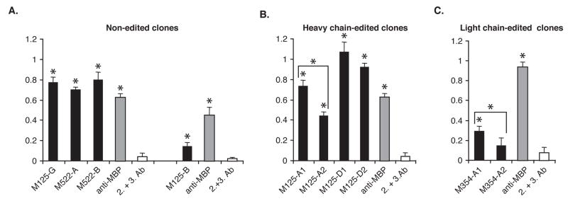
Panel A. MBP reactivity of the non-edited Fab clones (black bars) and a commercially available anti-MBP antibody (gray bars), or the secondary and tertiary antibodies alone (white bars). Panel B. MBP reactivity of the heavy chain edited Fab clones by ELISA. The receptor edited clone member M125-A2 had a significantly decreased reactivity to MBP compared to the non-edited clone member M125-A1. The non-edited and receptor edited clone members of M125-D had similar reactivity to MBP by ELISA. Panel C. MBP reactivity of the light chain edited clone M354-A2 had a significantly decreased reactivity to MBP in comparison to the non-edited clone member, M354-A1. In all assays, the ELISA plates were coated with whole human MBP (1 ug/mL) and ELISA was performed as described in Materials and Methods. Y-axis readout (color intensity) was determined at O.D. 405. Data shown represent the mean values for triplicate samples. Asterix (*) indicate Fabs that reacted to MBP statistically greater than background as assessed by student t-test (p≤0.05). This ELISA is representative of 6 independent experiments.
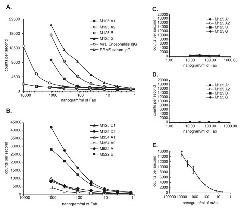
Plates were coated with either MBP (Panels A and B), Histone H1 (Panel C), or Lysozyme (Panel D) which are the control antigens with similar pI to MBP. Panel E. MBP binding of the anti-MBP antibody used throughout this study by DELFIA. A serum sample from a patient with confirmed Viral Encephalomyelitis (with high anti-MBP reactivity) and a serum sample from a patient with RRMS (with low anti-MBP reactivity) were included in the assay as internal controls and are shown in Panel A. M125-A1, B, D2, G and M522-B bind MBP with high titer, M125-D1, M522-A, M354-A1 and A2 bind MBP with low titer, and M125-A2 had negligible MBP reactivity.
References
-
- Antel J. Multiple sclerosis - emerging concepts of disease pathogenesis. J Neuroimmunol. 1999;98:45–48. - PubMed
-
- Archelos JJ, Storch MK, Hartung HP. The role of B cells and autoantibodies in multiple sclerosis. Annu Neurol. 2000;47:694–706. - PubMed
-
- Berger T, Rubner P, Schautzer F, Egg R, Ulmer H, Mayringer I, Dilitz E, Deisenhammer F, Reindl M. Antimyelin antibodies as a predictor of clinically definite multiple sclerosis after a first demyelinating event. N Engl J Med. 2003;349:139–145. - PubMed
Publication types
MeSH terms
Substances
Grants and funding
LinkOut - more resources
Full Text Sources
Other Literature Sources
Miscellaneous
