. 2007 Jul;33(7):1149-66.
doi: 10.1016/j.ultrasmedbio.2007.01.007. Epub 2007 Apr 23.

The impact of physiological motion on tissue tracking during radiation force imaging

Affiliations

The impact of physiological motion on tissue tracking during radiation force imaging

Brian J Fahey et al. Ultrasound Med Biol. 2007 Jul.

Abstract

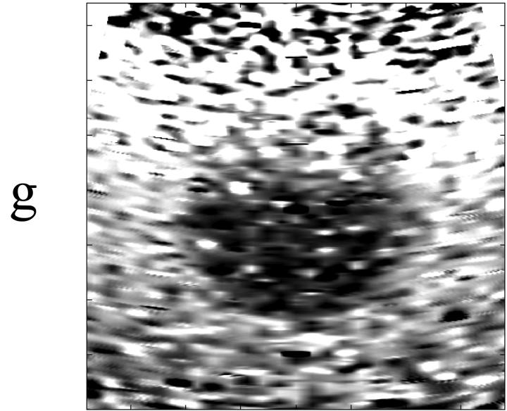
The effect of physiological motion on the quality of radiation force elasticity images has been investigated. Experimental studies and simulated images were used to investigate the impact of motion effects on image quality metrics over a range of clinically realistic velocity and acceleration magnitudes. Evaluation criteria included motion filter effectiveness, image signal-to-noise ratio (SNR) and the contrast-to-noise ratio (CNR) of a stiff inclusion embedded in a homogeneous background material. Two transmit frequencies (2.5 and 4.4 MHz) were analyzed and contrasted in terms of image quality over a range of target motions. Results indicate that situations may exist where liver and cardiac motion magnitudes lead to poor image quality, but optimized transducer orientations may help suppress motion artifacts if some a priori information concerning target motion characteristics is known. In the presence of significant target motion, utilizing a lower transmit frequency can improve SNR and CNR in elasticity images.


Figures

Figure 1
Figure 1
Functionality of linear motion filter. (a) shows the tracked displacement when ARFI data are acquired in the presence of 4 mm/s axial target motion. Assuming target region has completely recovered from radiation force excitation after 2.3 ms (arrow), we use the displacement tracked at this time (circled data point) to estimate the displacement profile attributable to transducer and/or target motion (dashed line). In (b) the original tracked displacement profile (squares) is shown, as well as the radiation force displacement profile (circles) recovered by subtracting the dashed profile in (a) from the original displacement estimate. (c) shows performance of filter when data are acquired in the presence of a 5 mm/s2 target acceleration.
Figure 1
Figure 1
Functionality of linear motion filter. (a) shows the tracked displacement when ARFI data are acquired in the presence of 4 mm/s axial target motion. Assuming target region has completely recovered from radiation force excitation after 2.3 ms (arrow), we use the displacement tracked at this time (circled data point) to estimate the displacement profile attributable to transducer and/or target motion (dashed line). In (b) the original tracked displacement profile (squares) is shown, as well as the radiation force displacement profile (circles) recovered by subtracting the dashed profile in (a) from the original displacement estimate. (c) shows performance of filter when data are acquired in the presence of a 5 mm/s2 target acceleration.
Figure 2
Figure 2
Curvilinear scanning geometry and associated coordinate terminology used in this document.
Figure 3
Figure 3
Qualitative validation of the liver motion model. (a) and (b) show normalized reference (stationary) experimental and simulated ARFI images, respectively. (c) and (d) show normalized experimental and simulated ARFI images acquired in the presence of 5 mm/s lateral target motion. Enclosed region in (b) corresponds to data provided in Table 2.
Figure 3
Figure 3
Qualitative validation of the liver motion model. (a) and (b) show normalized reference (stationary) experimental and simulated ARFI images, respectively. (c) and (d) show normalized experimental and simulated ARFI images acquired in the presence of 5 mm/s lateral target motion. Enclosed region in (b) corresponds to data provided in Table 2.
Figure 4
Figure 4
Motion filter effectiveness in the presence of target acceleration. (a) and (d) show images in the presence of 10 and 40 cm/s2 axial acceleration, respectively. (b) and (e) show images for 100 and 300 cm/s2 lateral accelerations, and (c) and (f) show images with 100 and 300 cm/s2 accelerations in the elevation dimension. All phantom targets were stationary at the time of radiation force application (t = 0). The ideal motion filter output is shown in Fig. 3(b). Region outlined in (a) corresponds to statistical analysis shown in Table 1.
Figure 4
Figure 4
Motion filter effectiveness in the presence of target acceleration. (a) and (d) show images in the presence of 10 and 40 cm/s2 axial acceleration, respectively. (b) and (e) show images for 100 and 300 cm/s2 lateral accelerations, and (c) and (f) show images with 100 and 300 cm/s2 accelerations in the elevation dimension. All phantom targets were stationary at the time of radiation force application (t = 0). The ideal motion filter output is shown in Fig. 3(b). Region outlined in (a) corresponds to statistical analysis shown in Table 1.
Figure 5
Figure 5
Effect of initial target velocity on motion filter performance in the presence of target acceleration. (a) shows a filtered image with an initial axial velocity of 2 cm/s and an axial acceleration of 10 cm/s2. (b) shows a filtered image with an initial lateral velocity of 2 cm/s and a lateral acceleration of 100 cm/s2.
Figure 5
Figure 5
Effect of initial target velocity on motion filter performance in the presence of target acceleration. (a) shows a filtered image with an initial axial velocity of 2 cm/s and an axial acceleration of 10 cm/s2. (b) shows a filtered image with an initial lateral velocity of 2 cm/s and a lateral acceleration of 100 cm/s2.
Figure 6
Figure 6
Effect of target velocity on image quality parameters with a 2.5 MHz transmit frequency. (a) shows results for normalized cross-correlation coefficient, (b) shows results for jitter. Results shown are for a scan line near the left-hand side of the image (-12°). Error bars represent ±1 standard deviation from the mean.
Figure 6
Figure 6
Effect of target velocity on image quality parameters with a 2.5 MHz transmit frequency. (a) shows results for normalized cross-correlation coefficient, (b) shows results for jitter. Results shown are for a scan line near the left-hand side of the image (-12°). Error bars represent ±1 standard deviation from the mean.
Figure 7
Figure 7
ARFI image SNR vs. time of displacement estimate. Shown are SNR values for the stationary phantom, a phantom with a 5 cm/s lateral velocity, and a phantom with a 10 cm/s elevation velocity. All times are relative to the end of the radiation force pulse. SNR values were calculated in the enclosed region shown in Fig. 3(b).
Figure 8
Figure 8
Comparison of ARFI images of a simulated inclusion phantom at two frequencies. Top row shows images with a transmit frequency of 2.5 MHz, bottom row with transmit frequency of 4.4 MHz. Shown from left to right are images acquired in the presence of (a and e) no motion, (b and f) 10 cm/s axial target velocity, (c and g) 10 cm/s elevation target velocity, and (d and h) 5 cm/s lateral target velocity. Dotted-line boxes in (a) show regions in all images used for CNR calculations. All images shown are motion filtered. Spatial scale of images is shown in (e).
Figure 8
Figure 8
Comparison of ARFI images of a simulated inclusion phantom at two frequencies. Top row shows images with a transmit frequency of 2.5 MHz, bottom row with transmit frequency of 4.4 MHz. Shown from left to right are images acquired in the presence of (a and e) no motion, (b and f) 10 cm/s axial target velocity, (c and g) 10 cm/s elevation target velocity, and (d and h) 5 cm/s lateral target velocity. Dotted-line boxes in (a) show regions in all images used for CNR calculations. All images shown are motion filtered. Spatial scale of images is shown in (e).

References

    1. Anderson W, Kissane J. Pathology. 9th edn. Mosby; St. Louis, MO: 1953.
    1. Bercoff J, Tanter M, Fink M. Supersonic shear imaging: a new technique for soft tissue elasticity mapping. IEEE Trans Ultrason Ferroelectr Freq Control. 2004;51(4):396–409. - PubMed
    1. Cespedes I, Ophir J, Alam S. The combined effect of signal decorrelation and random noise on the variance of time delay estimation. IEEE Trans. Ultrason., Ferroelec., Freq. Contr. 1997;44(1):220–225. - PubMed
    1. Chandrasekhar R, Ophir J, Krouskop T, Ophir K. Elastographic image quality vs. tissue motion in vivo. Ultrasound Med Biol. 2006;32(6):847–855. - PubMed
    1. Cheung MM, Smallhorn JF, McCrindle BW, Van Arsdell GS, Redington AN. Noninvasive assessment of ventricular force-frequency relations in the univentricular circulation by tissue doppler echocardiography: a novel method of assessing myocardial performance in congenital heart disease. Heart. 2005;91(10):1338–1342. - PMC - PubMed

Publication types