. 2007 Jul 1;396(1):93-107.
doi: 10.1016/j.gene.2007.02.031. Epub 2007 Mar 23.

Radial spoke protein 44 (human meichroacidin) is an axonemal alloantigen of sperm and cilia

Affiliations

Radial spoke protein 44 (human meichroacidin) is an axonemal alloantigen of sperm and cilia

Jagathpala Shetty et al. Gene. .

Abstract

To identify novel sperm alloantigens relevant to immune infertility, sera from infertile men containing antisperm antibodies (ASA) were employed on 2-D immunoblots of human sperm proteins. An immunoreactive protein spot (MW: 44 kDa, pI: 4.5) was microsequenced and the related cDNA was cloned yielding a 309 amino acid sequence corresponding to a gene currently annotated in Genbank as TSGA2 homolog (mouse) to signify 'testis specific gene A2'. In Genbank the protein deduced from this gene is currently named human meichroacidin, an orthologue of meichroacidin previously identified in mouse spermatocytes. Human TSGA2 mapped to chromosome 21q22.3. Human meichroacidin (hMCA) contained a single potential tyrosine phosphorylation site and five casein kinase phosporylation motifs. The N-terminus contained a Membrane Occupation Recognition Nexus (MORN) motif found in the lipid kinase-phosphatidylinositol 4-phosphate 5-kinase (PIP5K) family and junctophilins. However hMCA lacked the characteristic kinase homology domain of PIP5K. Northern blot analysis revealed 1.5 kb hMCA transcripts in testis and trachea with lower levels in thyroid and spinal cord. A semi-quantitative reverse transcription polymerase chain reaction (RT-PCR) analysis demonstrated occurrence of the mRNA messages in all the ciliated tissues tested with highest levels of messages in testis and trachea. Western blot analysis showed the presence of hMCA protein in brain, thyroid and trachea at the identical mass, 44 kDa, as in human testis. However, this immunoreactive pattern differed from that of sperm in which a 38 kDa form was also evident suggesting that hMCA undergoes proteolytic processing. In human testis, hMCA localized to the tails of developing spermatids and did not localize to the nucleus of either spermatocytes or spermatids. EM immunocytochemistry localized hMCA within the radial spokes of the axonemal complex of the sperm flagellum, and immunofluorescence studies revealed h-meichroacidin in the cilia of epithelial cells in the trachea and ependyma. Bioinformatic identification of orthologues of meichroacidin in several lower organisms including ciliates and flagellates suggest the protein plays a role in flagellar motility across phyla. We propose the term radial spoke protein 44 as an accurate designation, preferable to human meichroacidin because it denotes the restricted localization of the protein to the radial spokes of the axonemes of both sperm and cilia. Further, since the human gene is expressed in brain, thyroid, trachea and lung in addition to testis, we suggest that the gene name be changed from TSGA2 [testis specific gene A2] to radial spoke protein 44 [RSP44].


Figures
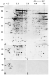
Fig. 1
Fig. 1
Identification of alloantigenic RSP44 (h-meichroacidin) spot by 2-D gel electrophoresis. A. Silver stained 2-D gel of Celis (Celis et al., 1992) extract of human sperm proteins showing the locations of h-meichroacidin (MA1, MA2 and MA3). The boxed area denotes the region of the gel shown in B-E. B: Sequential probing of a blot of sperm proteins using sera from 5 antisperm antibody positive infertile patients recognized 2 spots corresponding to h-meichroacidin, MA1 and MA2. C: Immunoblotting with 5 antisperm antibody negative, fertile subjects' sera did not recognize these spots. D: 2-D blot of human sperm proteins probed with rat serum against purified rec RSP44. The region corresponding to the originally microsequenced spots bound the antibody. E: Preimmune control for D.
Fig. 2
Fig. 2
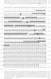
Fig 2: AThe nucleotide and deduced amino acid sequences of RSP44 cDNA. The deduced amino acid sequence is shown below the cDNA sequence. The numbers on the left refer to the amino acid sequence; numbers on the right refer to the nucleotide sequence. The consensus ATG of the ORF is shown in bold. The termination codon (TAA) is marked with an asterisk. The partial 5' and 3' untranslated regions are shown in italics. The polyadenylation signal is underlined with a thin line. The calculated molecular mass and pI of the predicted protein were 35.1 and 4.5 respectively. The sequences shaded in gray indicate the tryptic peptides obtained by microsequencing the gel spots noted in Fig. 1. The 7 MORN repeat sequences are underlined with a dark line and are numbered. The glutamic acid rich region is boxed. Fig. 2 B: Amino acid sequence alignment of RSP44 and mouse MCA. The identical amino acids are shaded in dark background with white letters and the similar amino acids are shaded in gray background with dark letters.
Fig. 2
Fig. 2
Fig 2: AThe nucleotide and deduced amino acid sequences of RSP44 cDNA. The deduced amino acid sequence is shown below the cDNA sequence. The numbers on the left refer to the amino acid sequence; numbers on the right refer to the nucleotide sequence. The consensus ATG of the ORF is shown in bold. The termination codon (TAA) is marked with an asterisk. The partial 5' and 3' untranslated regions are shown in italics. The polyadenylation signal is underlined with a thin line. The calculated molecular mass and pI of the predicted protein were 35.1 and 4.5 respectively. The sequences shaded in gray indicate the tryptic peptides obtained by microsequencing the gel spots noted in Fig. 1. The 7 MORN repeat sequences are underlined with a dark line and are numbered. The glutamic acid rich region is boxed. Fig. 2 B: Amino acid sequence alignment of RSP44 and mouse MCA. The identical amino acids are shaded in dark background with white letters and the similar amino acids are shaded in gray background with dark letters.
Fig. 3
Fig. 3
Northern blot analysis of RSP44. Two northern blots (Clontech) containing poly (A)+ mRNA from human tissues (2 μg per lane) were hybridized with radiolabeled RSP44 cDNA and exposed for 96 h. RNA markers are indicated on the left. RSP44 transcripts of ∼1.8 kb were clearly detected in testis and trachea. A low amount of mRNA expression was also detected in thyroid and spinal cord. The arrow indicates the reactivity to a testis mRNA that was spotted as a positive control.
Fig. 4
Fig. 4
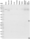
A multiple tissue Western blot analysis of RSP44. A: Protein samples were extracted from adult human tissues with NP40 and examined by Western blot analysis using rat anti-rec RSP44 polyclonal antibody. Protein samples (∼30μg/lane) from the brain, spinal cord, thyroid, liver, trachea, spleen, testis, uncapacitated sperm, and capacitated sperm were loaded. Expression of the protein in sperm was characterized by the appearance of 44 and 38 k Da bands in both un-capacitated and capacitated sperm. Brain and testis showed only the 44 k Da form of RSP44 with an intensity similar to that of sperm MCA. Thyroid and trachea also showed a lower level of expression of the higher molecular weight form of MCA. Spinal cord showed a pattern of expression similar to that of sperm, although the lower molecular weight form appeared to be of lower mass than that of sperm. Liver and spleen did not show any signal indicating the absence of protein in these tissues. B. Pre-immune control for A showed no reactivity at the expected molecular weight range for RSP44, although a low intensity background signal was observed at ∼25 k Da in both A and B.
Fig. 5
Fig. 5
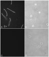
Indirect immunofluorescence of swim-up, permeabilized human sperm, including immunofluorescence staining for meichroacidin (A and C) and phase contrast pictures (B and D). The sperm were permeabilized with 0.1% saponin before probing with serum. The immune serum (A) stained the entire sperm tail including middle piece, principal piece and end piece. No immunostaining was observed with preimmune serum (C).
Fig. 6
Fig. 6
Immunoelectron microscopy of human spermatozoa: A and B: cross sections of the mid-piece of a human sperm tail, stained with anti-RSP44 antibody. Inside the layer of outer dense fibers, note the gold particles in the axoneme between the central microtules and the outer doublet of microtubules corresponding to the region occupied by the radial spokes. C: A longitudinal section of the principal piece of a human spermatozoon stained with anti-RSP44 serum. Note the gold particles associated with axonemal structure of the sperm tail. D: A schematic diagram of a cross section of the mid-piece of a human sperm illustrating the ultrastructural details of the axoneme (drawn based on Fawcett, 1975). E and F: A cross section (E) and a longitudinal section (F) of the principal piece of the human spermatozoon stained with pre-immune serum reveals absence of gold particles. FS: Fibrous sheath, ODF: outer dense fiber, Axo: Axoneme, Mito: Mitochondrion.
Fig. 7
Fig. 7
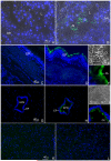
Indirect immmunofluorescent staining of sections of human testis (A and B), trachea (C-F) spinal cord (G-J) and liver (K and L). Sections were probed with either rat anti-meichroacidn sera (B, D and H) or pre-immune sera (A, C and G) and prepared for indirect immunofluorescence staining. The same sections were counterstained with DAPI II for nuclear staining. The antibody mainly stained the tails of the testicular spermatozoa (ts) in the lumen (L) of the seminiferous tubule. In the trachea, a region at the luminal surface of the epithelium corresponding to the location of ciliary structures was stained by the antibody (D and F). Similarly the cilia of the ependymal epithelium lining the central canal of the spinal cord were stained with the antibody (H and J). Figures E and I represent phase contrast images showing magnified views of a portion of the ciliated epithelium of the trachea and ependyma respectively, F and J depict the corresponding images stained with the RSP44 antibody showing staining of the cilia (CL). The liver section did not show any immunoreactivity when stained with the immune serum (L) and was comparable to the pre-immune control (K).
Fig. 8
Fig. 8
Semi-quantitative Reverse transcription polymerase chain reaction using total RNA from different ciliated tissues (A). Products generated by primer sets for hMCA were normalized against G3-PDH mRNA levels in the same set of samples. B) The relative densities of each band is plotted to show the level of message in different tissues.
Fig. 9
Fig. 9
The antisperm antibody positive infertile serum that led to the identification of RSP44 also reacted with the recombinant protein. RSP44 with a C-terminal histidine tag was expressed in E.coli using the pET28b plasmid, induced by addition of 1.0 mM IPTG, and purified by nickel ion affinity chromatography followed by Prep cell. A) Bacterial extracts and purified rec RSP44 stained with Coomassie blue. B) Immunoblot of bacterial extracts and purified rec RSP44 with anti-His antibody. C) Immunoblot of purified rec RSP44 with rat antisera against rec RSP44. D) Immunoblot of purified rec RSP44 with the infertile male serum which was originally used to identify RSP44 in a 2-D immunoblot of human sperm proteins. The infertile serum recognized the recombinant protein showing a profile similar to that shown in panels A, B and C, with a major band at ∼44k Da and few lower forms. The fertile serum (D) did not result in any signal with the rec RSP44 confirming that reactivity of the infertile serum is specific.

Similar articles

Cited by

References

    1. Anderson RA, Boronenkov IV, Doughman SD, Kunz J, Loijens JC. Phosphatidylinositol phosphate kinases, a multifaceted family of signaling enzymes. J Biol Chem. 1999;274:9907–9910. - PubMed
    1. Bouchard P, Cosson MP. Molecular basis of sperm movement. Ann. Endocrinol. (Paris) 1981;42:398–406. - PubMed
    1. Bronson RA, Cooper GW, Rosenfeld DL. Correlation between regional specificity of antisperm antibodies to the spermatozoan surface and complement-mediated sperm immobilization. Am. J. Reprod. Immunol. 1982;2:222–224. - PubMed
    1. Bronson RA, Tung KSK. Human sperm antibodies and their detection. In: Rose NR, de Macario ER, editors. Am. Soc. for Microbiol. 4th edition Manual of Clinical Laboratory Immunology; Washington, DC: 1992. pp. 1013–1017.
    1. Caron CP, Saling PM. Sperm antigens and immunological interference of fertilization. In: Wassarman PM, editor. Elements of Mammalian Fertilization. CRC Press; Boca Raton, FL: 1991. pp. 147–176.

Publication types

MeSH terms