Review
. 2007 Jun;1772(6):629-44.
doi: 10.1016/j.bbadis.2007.02.011. Epub 2007 Mar 2.

The cellular prion protein (PrP(C)): its physiological function and role in disease

Affiliations
Review

The cellular prion protein (PrP(C)): its physiological function and role in disease

Laura Westergard et al. Biochim Biophys Acta. 2007 Jun.

Abstract

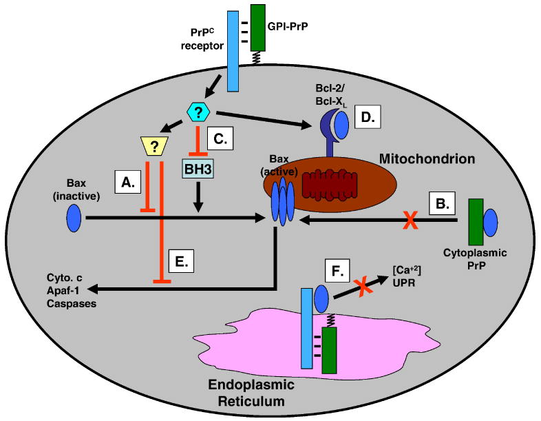
Prion diseases are caused by conversion of a normal cell-surface glycoprotein (PrP(C)) into a conformationally altered isoform (PrP(Sc)) that is infectious in the absence of nucleic acid. Although a great deal has been learned about PrP(Sc) and its role in prion propagation, much less is known about the physiological function of PrP(C). In this review, we will summarize some of the major proposed functions for PrP(C), including protection against apoptotic and oxidative stress, cellular uptake or binding of copper ions, transmembrane signaling, formation and maintenance of synapses, and adhesion to the extracellular matrix. We will also outline how loss or subversion of the cytoprotective or neuronal survival activities of PrP(C) might contribute to the pathogenesis of prion diseases, and how similar mechanisms are probably operative in other neurodegenerative disorders.


Figures

FIGURE 1
FIGURE 1. Possible mechanisms for PrP suppression of Bax-induced apoptosis
PrPC may inhibit Bax-mediated apoptotic pathways at several different points, either by a direct interaction between the two proteins or by involvement of additional, intermediary proteins. PrPC on the cell surface (GPI-PrP) may bind to a putative transmembrane receptor, initiating a signal transduction cascade that culminates in inhibition of Bax mitochondrial translocation, conformational change, or oligomerization (A). Cytoplasmic forms of PrP may produce similar effects via a direct interaction with Bax (B). PrP may inhibit pro-apoptotic, BH3-only proteins (C), or enhance an interaction between Bax and anti-apoptotic, multi-domain proteins such as Bcl-2 and Bcl-XL (D). PrP may suppress downstream events in the Bax pathway, such as cytochrome c (cyto. c) release, or activation of Apaf-1 and caspases (E). Finally, PrP in the ER may alter Bax function in this organelle, via effects on intracellular calcium and the unfolded protein response (UPR) (F).
FIGURE 2
FIGURE 2. PrP targeted to the secretory pathway protects yeast against Bax-induced cell death
(A) S. cerevisiae expressing Bax from a galactose-inducible promoter were transformed with empty vector, or with vector constitutively expressing either PrP or human Bax-inhibitor 1 (BI-1) (a positive control protein). The PrP construct was engineered to allow expression in the secretory pathway [44]. Three independent transformants (a-c) were spotted in serial 5-fold dilutions (left to right) on glucose or galactose plates and allowed to grow for 3 or 6 days, respectively. Co-expression of PrP allows yeast to grow on galactose medium (i.e., under conditions where Bax synthesis is induced). (B) Quantitation of the protective effects of PrP and BI-1. Yeast transformed as in (A) were plated onto glucose or galactose plates, and the number of colonies counted. Results are expressed as the number of colonies on the galactose plates as a % of those on the glucose plates. PrP is even more potent than BI-1 in restoring growth in the presence of Bax. (C) Lysates prepared from three independent yeast transformants (as described in A) were subjected to Western blotting using anti-PrP (upper panel, lanes 1-7), anti-HA to detect BI-1 (upper panel, lanes 8-10), or anti-Bax (lower panel, lanes 1-10). Lane 1 shows yeast that carry the empty vectors used for Bax and PrP/BI-1 expression. Neither PrP nor BI-1 affect expression levels of Bax. (D) Deletion analysis to determine which domains of PrP are required for its ability to protect yeast from Bax-induced cell death. Yeast expressing Bax from a galactose-inducible promoter were transformed with plasmids encoding wild-type (WT) PrP, or the indicated deletion constructs. Growth on glucose and galactose plates was assessed as in panels A and B in order to score PrP rescue activity. The green boxes indicate the hybrid signal sequence used to target PrP to the secretory pathway [44], and the blue boxes indicate the octapeptide repeats. Data are from Li and Harris [43].
FIGURE 3
FIGURE 3. Models for the cellular toxicity of PrPSc
(A) Toxic gain-of-function mechanism. PrPSc (or PrPtoxic, a pathogenic intermediate) possesses a novel neurotoxic activity that is independent of the normal function of PrPC. (B) Loss-of-function mechanism. PrPC possesses a normal, physiological activity, in this case neuroprotection, that is lost upon conversion to PrPSc. (C) Subversion-of-function mechanism. The normal, neuroprotective activity of PrPC is subverted by binding to PrPSc (or PrPtoxic). Cross-hatching of the rectangle representing PrPC indicates a change in its signaling properties such that a neurotoxic rather than a neuroprotective signal is delivered. Taken from Harris and True [148].

References

    1. Büeler H, Fischer M, Lang Y, Fluethmann H, Lipp HP, DeArmond SJ, Prusiner SB, Aguet M, Weissmann C. Normal development and behavior of mice lacking the neuronal cell-surface PrP protein. Nature. 1992;356:577–582. - PubMed
    1. Manson JC, Clarke AR, Hooper ML, Aitchison L, McConnell I, Hope J. 129/Ola mice carrying a null mutation in PrP that abolishes mRNA production are developmentally normal. Mol Neurobiol. 1994;8:121–127. - PubMed
    1. Mallucci GR, Ratte S, Asante EA, Linehan J, Gowland I, Jefferys JG, Collinge J. Post-natal knockout of prion protein alters hippocampal CA1 properties, but does not result in neurodegeneration. EMBO J. 2002;21:202–210. - PMC - PubMed
    1. Rivera-Milla E, Oidtmann B, Panagiotidis CH, Baier M, Sklaviadis T, Hoffmann R, Zhou Y, Solis GP, Stuermer CA, Malaga-Trillo E. Disparate evolution of prion protein domains and the distinct origin of Doppel- and prion-related loci revealed by fish-to-mammal comparisons. FASEB J. 2006;20:317–319. - PubMed
    1. Manson J, West JD, Thomson V, McBride P, Kaufman MH, Hope J. The prion protein gene: a role in mouse embryogenesis? Development. 1992;115:117–122. - PubMed

Publication types