. 2007 Jul;27(13):4774-83.
doi: 10.1128/MCB.00283-07. Epub 2007 Apr 23.

Ubiquitination-induced conformational change within the deiodinase dimer is a switch regulating enzyme activity

Affiliations

Ubiquitination-induced conformational change within the deiodinase dimer is a switch regulating enzyme activity

G D Vivek Sagar et al. Mol Cell Biol. 2007 Jul.

Abstract

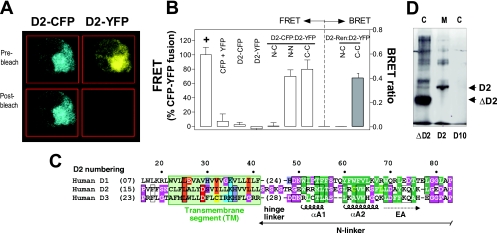
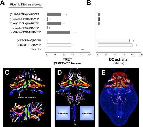
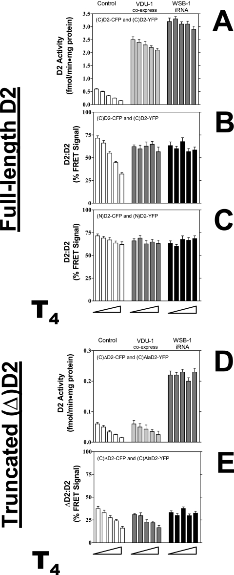
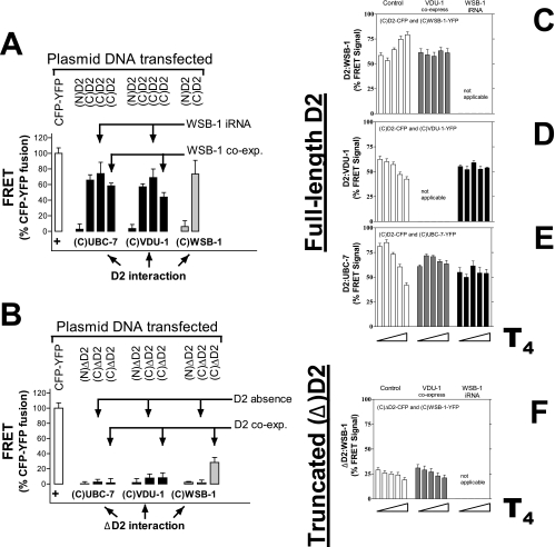
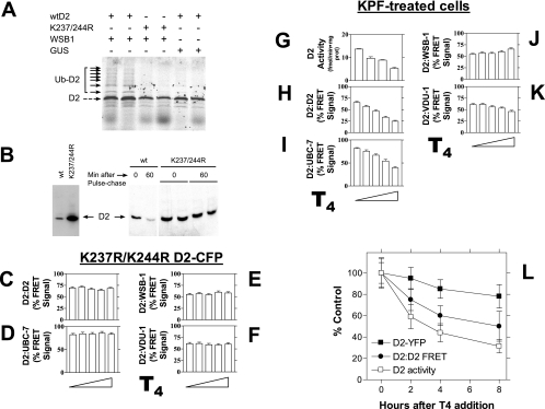
Ubiquitination is a critical posttranslational regulator of protein stability and/or subcellular localization. Here we show that ubiquitination can also regulate proteins by transiently inactivating enzymatic function through conformational change in a dimeric enzyme, which can be reversed upon deubiquitination. Our model system is the thyroid hormone-activating type 2 deiodinase (D2), an endoplasmic reticulum-resident type 1 integral membrane enzyme. D2 exists as a homodimer maintained by interacting surfaces at its transmembrane and globular cytosolic domains. The D2 dimer associates with the Hedgehog-inducible ubiquitin ligase WSB-1, the ubiquitin conjugase UBC-7, and VDU-1, a D2-specific deubiquitinase. Upon binding of T4, its natural substrate, D2 is ubiquitinated, which inactivates the enzyme by interfering with D2's globular interacting surfaces that are critical for dimerization and catalytic activity. This state of transient inactivity and change in dimer conformation persists until deubiquitination. The continuous association of D2 with this regulatory protein complex supports rapid cycles of deiodination, conjugation to ubiquitin, and enzyme reactivation by deubiquitination, allowing tight control of thyroid hormone action.


Figures

FIG. 1.
FIG. 1.
D2 holoenzyme is a homodimer. (A) Photomicrography of an individual HEK-293 cell transiently coexpressing D2-CFP and D2-YFP, pre- and post-YFP photobleaching. (B) Quantification of D2-D2 FRET in cells expressing D2-CFP and/or D2-YFP fused at the indicated termini of D2. The location of the chromophores in the D2 molecule is indicated by N (amino) or C (carboxyl) for CFP and YFP, respectively. Results were calculated as a percentage of the signal measured in cells expressing a CFP-YFP positive-control (+) fusion protein. In these positive-control cells, an approximately 20% increase in CFP fluorescence after YFP photobleaching is typically observed (data not shown). Also shown (right axis) is the D2-D2 BRET ratio in cells expressing D2-RLuc and D2-YFP. The location of the chromophores in the D2 molecule is indicated by N (amino) or C (carboxyl) for YFP and RLuc, respectively. Data are means ± standard deviations of at least 10 data points. (C) Partial sequence alignment of human D1, D2, and D3, denoting the transmembrane and N-linker regions (6). Hydrophobic residues are green, loop-forming residues are depicted by white letters on a pink background, and the neutral amino acid H is shown on a purple background; the putative transmembrane segment (20 aa) is shown on a light green background, with amino acids likely to be in tight contact in the dimer interface shown in orange. Red, acidic; blue, basic; yellow, cysteine. (D) Western blot analysis with anti-FLAG antibody of cytosol (C) or microsomal (M) subcellular fractions of cells transiently expressing a truncated D2 without the transmembrane domain (ΔD2), full-length D2 (D2), or empty D10 vector.
FIG. 2.
FIG. 2.
Globular interfaces mediate D2 dimerization and are critical for catalytic activity. (A) FRET signal in cells expressing the indicated D2 proteins fused to either CFP or YFP at the amino (N) or carboxyl (C) termini of the D2 molecule. AlaD2 is a full-length D2 molecule in which Sec133 was mutated to Ala. (B) D2 activity in cell sonicates shown in panel A. In panels A and B, data are means ± standard deviations of at least 10 data points. (C) Two orthogonal views of the modeled D2 dimer on the template of the crystal structure of human thioredoxin dimer. At the top, the twofold axis is vertical, and at the bottom, it is perpendicular to the figure. Secondary structures are colored as published elsewhere (6) and labeled accordingly. The putative structure of the iduronidase-like active site insertion has been modeled as a ββ secondary structure (βd1 and βd2) lying between β2 and αB. βd1 is colored green/light purple, βd2 is in in dark purple, and at the bottom of the dimer, the two symmetrical small βT structures (D2 G224, V225, A226; pink) are the counterparts of the canonical thioredoxin pairing. (D) 3D model of the D2-D2 dimer. N′-ter and C-ter indicate the N terminus of the thioredoxin fold head domain and the C terminus of D2, respectively. The S42-K76 connecting segment is rich in strongly hydrophobic amino acids (VILFMYW), which are shown in atomic detail. The N and C termini of this segment readily match the C and N termini of the transmembrane and globular domains, respectively. The buried dimeric interface for the full model is 2,170 Å2 (in the range observed for moderately strong dimers and slightly higher than those of many protein complexes [1]), that for the head is 1,343 Å2 (more than twice that for the thioredoxin dimer), that for the N-linker is 128 Å2, and that for the transmembrane segment is 705 Å2, confirming the respective roles of each part, particularly that of the transmembrane segments. A single large cavity (427 Å2) is created upon D2 dimerization at the level of the active site (Sec133), which is surrounded by S130, T132, Se133, P134, I161, D162, M215, N218, Y223, G224, V255, A226, E228, and their symmetric residues. (E) Visualization of the Russian-doll-shaped electrostatic field around the D2 dimer (the −1.8-kT/e gradient limit is colored red and the +1.8-kT/e gradient limit is blue).
FIG. 3.
FIG. 3.
Properties of D2-D2 dimer during catalysis. In all experiments cells were treated with 0, 0.1, 1, 5, or 10 μM T4 (from left to right) in 10% charcoal-stripped fetal bovine serum-containing medium for 4 h immediately prior to harvesting or FRET studies. (A) D2 activity in cells coexpressing full-length D2-CFP and D2-YFP; chromophores were placed at the carboxyl (C) terminus of D2; VDU-1 or WSB-1 interfering RNA (iRNA) plasmids were also used as indicated. (B) D2-D2 FRET signal in cells transfected as in panel A. (C) D2-D2 FRET signal in cells coexpressing D2-CFP and D2-YFP; chromophores were placed at the amino (N) terminus of D2. (D) Same as in panel A, except that ΔD2 was used. (E) ΔD2-D2 FRET signal in cells transfected as in panel D. Data are means ± standard deviations of at least 10 data points.
FIG. 4.
FIG. 4.
D2 interaction with UBC-7, WSB-1, and VDU-1. (A) FRET studies in HEK-293 cells transiently coexpressing D2-CFP and UBC-7-YFP, WSB-1-YFP, or VDU-1-YFP. As indicated, some studies were performed in cells with WSB-1 knockdown (interfering RNA [iRNA]) or coexpressing WSB-1. The positions of the chromophores in the D2 molecule are indicated as amino (N) or carboxyl (C) termini. (B) Same as panel A, except that the interactions of ΔD2 with UBC-7, VDU-1, or WSB-1 were studied in the absence of or during full-length D2 coexpression. In the experiments for panels C to F cells were treated with 0, 0.1, 1, 5, or 10 μM T4 (from left to right) in 10% charcoal-stripped fetal bovine serum-containing medium for 4 h immediately prior to FRET studies. (C to E) D2 interaction with WSB-1 (C), with VDU-1 (D), and with UBC-7 (E), with or without VDU-1 or WSB-1. (F) Same as in panel C, except that ΔD2 was used. Data are means ± standard deviations of at least 10 data points.
FIG. 5.
FIG. 5.
Mechanism of D2 ubiquitination. (A) A 35S-labeled D2 or K237R/K244R D2 mutant was used in the in vitro ubiquination assay in a reticulocyte lysate system containing 10 μM ubiquitin aldehyde, energy solution, and 30 μM ubiquitin as previously described (16). Bacterially expressed WSB-1 or Arabidopsis thaliana β-glucoronidase (GUS) was added as indicated after normalization by Coomassie blue stain SDS-PAGE. Ubiquitinated D2 was immunoprecipitated using D2 antiserum (IP α-D2), and the pellets were resolved by SDS-PAGE. The black arrows indicate the ubiquitin-35S-D2 conjugates (Ub-35S-D2). wt, wild type. (B) Immunoprecipitation of 35S-labeled D2 or K237R/K244R D2 mutant using anti-FLAG after a 60-min pulse-chase. In the experiments shown in panels C to F and H to K, cells were treated with 0, 0.1, 1, 5, or 10 μM T4 (from left to right) in 10% charcoal-stripped fetal bovine serum-containing medium for 4 h immediately prior to harvesting or FRET studies. (C to F) K237R/K244R D2-CFP mutant was used. FRET signals in cells expressing the indicated proteins are shown: D2-D2 (C), D2-UBC-7 (D), D2-WSB-1 (E), and D2-VDU-1 (F). (G and H) D2 activity (G) and D2-D2 FRET signal (H) in cells coexpressing full-length D2-CFP and D2-YFP and pretreated with 40 μM kaempferol (KPF) for 12 h. For D2 activity (G), treatments were 0, 0.1, 1, or 5 mM T4. (I to K) Under similar conditions the FRET signals between D2 and UBC-7 (I), D2 and WSB-1 (J), and D2 and VDU-1 (K) were also obtained. (L) Same as in panels G and H except that cells were treated with 10 μM T4 for the indicated times and D2-YFP signal was also measured. Data are means ± standard deviations of at least 10 data points.

References

    1. Bahadur, R. P., P. Chakrabarti, F. Rodier, and J. Janin. 2004. A dissection of specific and non-specific protein-protein interfaces. J. Mol. Biol. 336:943-955. - PubMed
    1. Baqui, M., D. Botero, B. Gereben, C. Curcio, J. W. Harney, D. Salvatore, K. Sorimachi, P. R. Larsen, and A. C. Bianco. 2002. Human type 3 iodothyronine selenodeiodinase is located in the plasma membrane and undergoes rapid internalization to endosomes. J. Biol. Chem. 4:4. - PubMed
    1. Baqui, M. M., B. Gereben, J. W. Harney, P. R. Larsen, and A. C. Bianco. 2000. Distinct subcellular localization of transiently expressed types 1 and 2 iodothyronine deiodinases as determined by immunofluorescence confocal microscopy. Endocrinology 141:4309-4312. - PubMed
    1. Berry, M. J., A. L. Maia, J. D. Kieffer, J. W. Harney, and P. R. Larsen. 1992. Substitution of cysteine for selenocysteine in type I iodothyronine deiodinase reduces the catalytic efficiency of the protein but enhances its translation. Endocrinology 131:1848-1852. - PubMed
    1. Bianco, A. C., and B. W. Kim. 2006. Deiodinases: implications of the local control of thyroid hormone action. J. Clin. Investig. 116:2571-2579. - PMC - PubMed

Publication types