. 2007 Jul;27(13):5047-54.
doi: 10.1128/MCB.02234-06. Epub 2007 Apr 23.

CpG hypomethylation in a large domain encompassing the embryonic beta-like globin genes in primitive erythrocytes

Affiliations

CpG hypomethylation in a large domain encompassing the embryonic beta-like globin genes in primitive erythrocytes

Mei Hsu et al. Mol Cell Biol. 2007 Jul.

Abstract

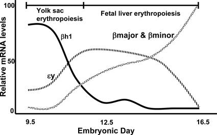
There is little evidence addressing the role of CpG methylation in transcriptional control of genes that do not contain CpG islands. This is reflected in the ongoing debate about whether CpG methylation merely suppresses retroelements or if it also plays a role in developmental and tissue-specific gene regulation. The genes of the beta-globin locus are an important model of mammalian developmental gene regulation and do not contain CpG islands. We have analyzed the methylation status of regions in the murine beta-like globin locus in uncultured primitive and definitive erythroblasts and other cultured primary and transformed cell types. A large ( approximately 20-kb) domain is hypomethylated only in primitive erythroid cells; it extends from the region just past the locus control region to before beta-major and encompasses the embryonic genes Ey, beta h1, and beta h0. Even retrotransposons in this region are hypomethylated in primitive erythroid cells. The existence of this large developmentally regulated domain of hypomethylation supports a mechanistic role for DNA methylation in developmental regulation of globin genes.


Figures

FIG. 1.
FIG. 1.
Changes in levels of β-like globin mRNAs during development. Note that the embryonic genes ɛy and βh1 are expressed in primitive erythroid cells generated in the yolk sac during early embryonic development, with transcripts undetectable by E16.5. Data are adapted from reference .
FIG. 2.
FIG. 2.
CpG methylation of murine β-like globin genes in cultured cells. Top, scaled map of the murine β-like globin locus. Vertical lines demarcate segments assayed. Open ovals, transcribed genes; filled ovals, pseudogenes; numbered filled ovals, DNase-hypersensitive sites in the LCR. Below, each horizontal row of blocks represents one bisulfite-treated clone of the indicated segment; vertical columns denote specific CpG sites. Open box, unmethylated; filled box, methylated.
FIG. 3.
FIG. 3.
Methylation status of CpGs surveyed in primary erythroid cells. (A) Top: isolation method and erythroid purity of samples. Bottom: representative FACScan for Ter-119 expression on Ter-119-sorted cells. (B) Organization of data similar to that in Fig. 2. Assayed segments are numbered for reference in the text. Sites marked with a # are Line elements, and the site with a Δ is a Sine element. The asterisks denote polymorphic CpGs, and unboxed sites in the column did not have that CpG.
FIG. 4.
FIG. 4.
Nongenic transcription within the hypomethylated domain. The map shows the sites of primers used to assay by RT-PCR. Numbered sites are at regions noted in Fig. 3B. G is a lane with genomic DNA; the + or − RT lanes denote whether reverse transcriptase was in the reaction mixture to produce cDNA and control for contamination with genomic DNA.
FIG. 5.
FIG. 5.
Model of interaction of methyl binding domain complexes around embryonic genes. Top, in primitive cells the lack of methylation across the domain blocks recruitment of MBD complexes. Bottom, methylation of the domain in definitive cells permits recruitment of MBD complexes, which contribute to gene silencing of the embryonic genes. Expressed genes are shown as hatched ovals.

Similar articles

Cited by

References

    1. Bulger, M., D. Schubeler, M. A. Bender, J. Hamilton, C. Farrell, M. R. C. Hardison, and M. Groudine. 2003. A complex chromatin landscape revealed by patterns of nuclease sensitivity and histone modification within the mouse β-globin locus. Mol. Cell. Biol. 23:5234-5244. - PMC - PubMed
    1. Charache, S., G. Dover, K. Smith, C. C. Talbot, M. Moyer, and S. Boyer. 1983. Treatment of sickle cell anemia with 5-azacytidine results in increased fetal hemoglobin production and is associated with nonrandom hypomethylation of DNA around the γδβ-globin gene complex. Proc. Natl. Acad. Sci. USA 80:4842-4846. - PMC - PubMed
    1. Constantoulakis, P., B. Josephson, L. Mangahas, T. Papayannopoulou, T. Enver, F. Costantini, and G. Stamatoyannopoulos. 1991. Locus control region-A gamma transgenic mice: a new model for studying the induction of fetal hemoglobin in the adult. Blood 77:1326-1333. - PubMed
    1. Costello, J., and P. Vertino. 2002. Methylation matters: a new spin on maspin. Nat. Genet. 31:123-124. - PubMed
    1. Ehrlich, M., M. A. Gama-Sosa, L. H. Huang, R. M. Midgett, K. C. Kuo, R. A. McCune, and C. Gehrke. 1982. Amount and distribution of 5-methylcytosine in human DNA from different types of tissues of cells. Nucleic Acids Res. 10:2709-2721. - PMC - PubMed

Publication types

LinkOut - more resources