. 2007 Jul;27(13):4617-25.
doi: 10.1128/MCB.00404-07. Epub 2007 Apr 23.

Yeast Rpb9 plays an important role in ubiquitylation and degradation of Rpb1 in response to UV-induced DNA damage

Affiliations

Yeast Rpb9 plays an important role in ubiquitylation and degradation of Rpb1 in response to UV-induced DNA damage

Xuefeng Chen et al. Mol Cell Biol. 2007 Jul.

Abstract

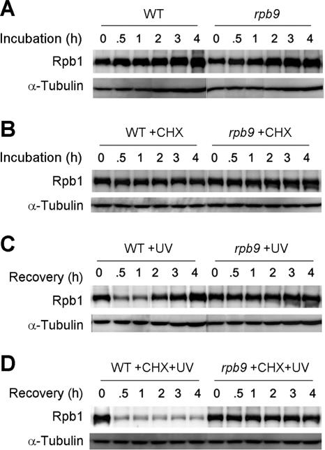
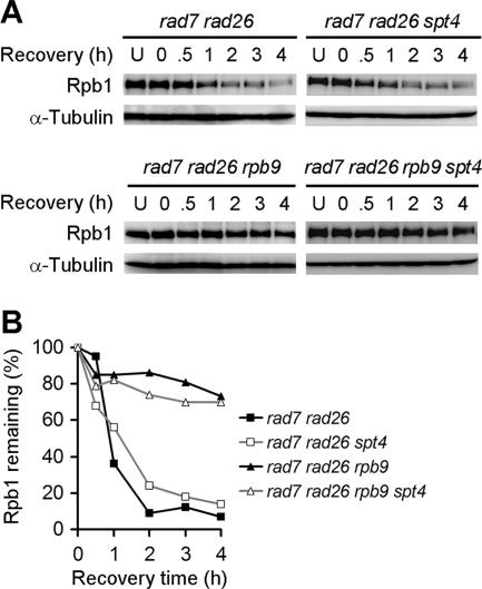
Rpb9, a nonessential subunit of RNA polymerase II (Pol II), has multiple transcription-related functions in Saccharomyces cerevisiae, including transcription elongation and transcription-coupled repair (TCR). Here we show that, in response to UV radiation, Rpb9 also functions in promoting ubiquitylation and degradation of Rpb1, the largest subunit of Pol II. This function of Rpb9 is not affected by any pathways of nucleotide excision repair, including TCR mediated by Rpb9 itself and by Rad26. Rpb9 is composed of three distinct domains: the N-terminal Zn1, the C-terminal Zn2, and the central linker. The Zn2 domain, which is dispensable for transcription elongation and TCR functions, is essential for Rpb9 to promote Rpb1 degradation, whereas the Zn1 and linker domains, which are essential for transcription elongation and TCR functions, play a subsidiary role in Rpb1 degradation. Coimmunoprecipitation analysis suggests that almost the full length of Rpb9 is required for a strong interaction with the core Pol II: deletion of the Zn2 domain causes dramatically weakened interaction, whereas deletion of Zn1 and the linker resulted in undetectable interaction. Furthermore, we show that Rpb1, rather than the whole Pol II complex, is degraded in response to UV radiation and that the degradation is primarily mediated by the 26S proteasome.


Figures

FIG. 1.
FIG. 1.
Selective degradation of Rpb1 in response to UV radiation. (A) Western blots showing the levels of Rpb1 and FLAG3-tagged (3×FLAG) Rpb2 and Rpb9 at different times following UV irradiation. Whole-cell extracts were prepared from aliquots of cultures taken at different recovery times following UV irradiation. Rpb1 and FLAG3-tagged Rpb2 and Rpb9 in the whole-cell extracts were probed with antibodies anti-Rpb1 (8WG16) and anti-FLAG (M2), respectively. Lanes U are unirradiated samples. α-Tubulin serves as an internal loading control. (B) Plots showing percentages of Rpb1 and FLAG3-tagged Rpb2 and Rpb9 remaining at different times following UV irradiation. The data shown in the plots were obtained by quantification of the Rpb1 and FLAG3-tagged Rpb2 and Rpb9 bands in the Western blots shown in panel A. The loading was normalized using the signal intensities of α-tubulin bands.
FIG. 2.
FIG. 2.
Rpb9 plays an important role in 26S proteasome-mediated Rpb1 degradation upon UV irradiation. Whole-cell extracts were prepared from aliquots of cultures taken at different recovery times following UV irradiation. Rpb1 levels in the extracts were probed with antibody 8WG16 on Western blots. Lanes U are unirradiated samples. α-Tubulin serves as an internal loading control. (A) Rpb1 levels in erg6 cells in the absence or presence of 50 μM of the 26S proteasome inhibitor MG132, which was added to the cultures 2 h prior to UV irradiation. (B) Rpb1 levels in rpb9 cells. (C) Plots showing percentages of Rpb1 remaining at different times following UV irradiation. The data shown in the plots were obtained by quantification of the Rpb1 bands in the Western blots shown in panels A and B. The loading was normalized using the signal intensities of α-tubulin bands.
FIG. 3.
FIG. 3.
Degradation of Rpb1 in rpb9 cells is impaired in response to UV radiation. Whole-cell extracts were prepared from aliquots of cultures taken at different incubation times. Rpb1 in the whole-cell extracts was probed with antibody 8WG16 on Western blots. α-Tubulin serves as an internal loading control. (A) Rpb1 levels in normally growing yeast cells. (B) Rpb1 levels in cells cultured in the presence of 500 μg/ml of CHX. (C) Rpb1 levels in cells at different times of recovery incubation following UV irradiation. (D) Rpb1 levels in cells at different times of recovery incubation in the presence of 500 μg/ml of CHX following UV irradiation. CHX was added to log-phase yeast cultures to a final concentration of 500 μg/ml. After 40 min of continued incubation, the cultures were directly irradiated with UV and recovered for different times.
FIG. 4.
FIG. 4.
Rpb9 promotes ubiquitylation of Rpb1 in response to UV radiation. (A) Western blot showing that slower-migrating bands, which reflect covalently modified Rpb1, are present in UV-irradiated WT cells but not rpb9 cells. Whole-cell extracts were prepared from aliquots of cultures taken at different recovery times following UV irradiation. Rpb1 in the extracts was probed with antibody H14 on the blot. As a significant degradation of Rpb1 occurred in WT cells during the recovery incubation, the amounts of loading were adjusted to give similar total signal intensities for samples of WT cells at different times of recovery incubation. (B) Ubiquitylated Rpb1 in immunoprecipitates from unirradiated and UV-irradiated cells. Cell lysates were prepared from unirradiated or UV-irradiated cells following 1 h of recovery incubation. Rpb1 was immunoprecipitated (IP) from the cell lysates using anti-Rpb1 antibody 8WG16. The immunoprecipitates were probed with antiubiquitin and 8WG16 antibodies on a Western blot. ub, ubiquitylated. (C) Western blot showing ubiquitylated proteins in whole-cell extracts from unirradiated and UV-irradiated cells. Proteins on the blot were probed with antiubiquitin antibody.
FIG. 5.
FIG. 5.
Role of Rpb9 domains in promoting Rpb1 degradation in response to UV radiation. (A) Sequence of Rpb9. The conserved cysteine residues for Zn2+ binding are underlined. The open bar underneath of the sequence marks the region that is essential for transcription elongation and TCR functions. (B) Western blots showing Rpb1 levels in rad16 rad26 rpb9 cells transformed with plasmids encoding full-length (1 to 122) or truncated (numbers indicate residues remaining) Rpb9 proteins. Whole-cell extracts were prepared from aliquots of cultures taken at different recovery times following UV irradiation. Rpb1 on the blots was probed with antibody 8WG16. Lanes U are unirradiated samples. α-Tubulin serves as an internal loading control.
FIG. 6.
FIG. 6.
Coimmunoprecipitation of truncated Rpb9 with core Pol II. Cell lysates were prepared from rad16 rad26 rpb9 cells transformed with plasmids encoding full-length (1 to 122) or truncated (numbers indicate residues remaining) FLAG3-tagged (3×FLAG) Rpb9 proteins. Protein complexes containing the FLAG3-tagged Rpb9 were immunoprecipitated (IP) with anti-FLAG antibody M2 or mock immunoprecipitated with mouse immunoglobulin G (IgG). Rpb1 and the tagged Rpb9 in the immunoprecipitates were probed with 8WG16 and anti-FLAG M2 antibodies, respectively, on Western blots. Cell lysates (immunoprecipitation inputs) were also probed with 8WG16 antibody on a Western blot to examine the intrinsic Rpb1 levels. α-Tubulin serves as an internal loading control.
FIG. 7.
FIG. 7.
The role of Rpb9 in promoting UV-induced degradation of Rpb1 is not affected by different NER pathways. Whole-cell extracts were prepared from aliquots of cultures taken at different recovery times following UV irradiation. Rpb1 levels in the extracts were probed with antibody 8WG16 on Western blots. Lanes U are unirradiated samples. α-Tubulin serves as an internal loading control. (A) Rpb1 levels in different rad deletion mutants. (B) Rpb1 levels in rpb9 cells with different rad deletions. (C) Rpb1 levels in def1 cells.
FIG. 8.
FIG. 8.
Reinstatement of TCR in otherwise TCR-deficient cells does not affect UV-induced Rpb1 degradation. (A) Whole-cell extracts were prepared from aliquots of cultures taken at different recovery times following UV irradiation. Rpb1 levels in the extracts were probed with antibody 8WG16 on Western blots. Lanes U are unirradiated samples. α-Tubulin serves as an internal loading control. (B) Plots showing percentages of Rpb1 remaining at different times following UV irradiation. The data shown were obtained by quantification of the Rpb1 bands in the Western blots shown in panel A. The loading was normalized using the signal intensities of α-tubulin bands.

Similar articles

Cited by

References

    1. Beaudenon, S. L., M. R. Huacani, G. Wang, D. P. McDonnell, and J. M. Huibregtse. 1999. Rsp5 ubiquitin-protein ligase mediates DNA damage-induced degradation of the large subunit of RNA polymerase II in Saccharomyces cerevisiae. Mol. Cell. Biol. 19:6972-6979. - PMC - PubMed
    1. Bergmann, E., and J. M. Egly. 2001. Trichothiodystrophy, a transcription syndrome. Trends Genet. 17:279-286. - PubMed
    1. Bregman, D. B., R. Halaban, A. J. van Gool, K. A. Henning, E. C. Friedberg, and S. L. Warren. 1996. UV-induced ubiquitination of RNA polymerase II: a novel modification deficient in Cockayne syndrome cells. Proc. Natl. Acad. Sci. USA 93:11586-11590. - PMC - PubMed
    1. Cramer, P. 2002. Multisubunit RNA polymerases. Curr. Opin. Struct. Biol. 12:89-97. - PubMed
    1. Cramer, P., D. A. Bushnell, and R. D. Kornberg. 2001. Structural basis of transcription: RNA polymerase II at 2.8 angstrom resolution. Science 292:1863-1876. - PubMed

Publication types

MeSH terms

LinkOut - more resources