. 2007 Jul;75(7):3361-72.
doi: 10.1128/IAI.01886-06. Epub 2007 Apr 23.

Toll-like receptor 2-dependent NF-kappaB activation is involved in nontypeable Haemophilus influenzae-induced monocyte chemotactic protein 1 up-regulation in the spiral ligament fibrocytes of the inner ear

Affiliations

Toll-like receptor 2-dependent NF-kappaB activation is involved in nontypeable Haemophilus influenzae-induced monocyte chemotactic protein 1 up-regulation in the spiral ligament fibrocytes of the inner ear

Sung K Moon et al. Infect Immun. 2007 Jul.

Abstract

Inner ear dysfunction secondary to chronic otitis media (OM), including high-frequency sensorineural hearing loss or vertigo, is not uncommon. Although chronic middle ear inflammation is believed to cause inner ear dysfunction by entry of OM pathogen components or cytokines from the middle ear into the inner ear, the underlying mechanisms are not well understood. Previously, we demonstrated that the spiral ligament fibrocyte (SLF) cell line up-regulates monocyte chemotactic protein 1 (MCP-1) expression after treatment with nontypeable Haemophilus influenzae (NTHI), one of the most common OM pathogens. We hypothesized that the SLF-derived MCP-1 plays a role in inner ear inflammation secondary to OM that is responsible for hearing loss and dizziness. The purpose of this study was to investigate the signaling pathway involved in NTHI-induced MCP-1 up-regulation in SLFs. Here we show for the first time that NTHI induces MCP-1 up-regulation in the SLFs via Toll-like receptor 2 (TLR2)-dependent activation of NF-kappaB. TLR2(-/-)- and MyD88(-/-)-derived SLFs revealed involvement of TLR2 and MyD88 in NTHI-induced MCP-1 up-regulation. Studies using chemical inhibitors and dominant-negative constructs demonstrated that it is mediated by the IkappaKbeta-dependent IkappaBalpha phosphorylation and NTHI-induced NF-kappaB nuclear translocation. Furthermore, we demonstrated that the binding of NF-kappaB to the enhancer region of MCP-1 is involved in this up-regulation. In addition, we have identified a potential NF-kappaB motif that is responsive and specific to certain NTHI molecules or ligands. Further studies are necessary to reveal specific ligands of NTHI that activate host receptors. These results may provide us with new therapeutic strategies for prevention of inner ear dysfunction secondary to chronic middle ear inflammation.


Figures
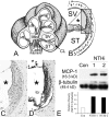
FIG. 1.
FIG. 1.
SLFs up-regulate MCP-1 upon exposure to NTHI. (A and B) Illustrations of the cochlear three-dimensional structure (A) and the cochlear lateral wall (B). Mo, modiolus; SV, scala vestibuli; ST, scala tympani; *, scala media; CoN, cochlear nerve; CL, cochlear lateral wall. (C and D) Immunolabeling of the cochlear lateral wall shows up-regulation of MCP-1 expression in the NTHI-treated group (D) compared to a control (C). Live NTHI was inoculated in the mouse middle ear, and the cochlea was dissected after 2 days. As a control, normal saline was inoculated with a same procedure. After decalcification and sectioning, immunolabeling was performed using a polyclonal anti-MCP-1 antibody. (E) Western blotting and its densitinogram show that SLFs up-regulate MCP-1 upon exposure to the NTHI lysate, compared to the PBS-treated control (Con). The spiral ligament cell line was treated with two different culture batches (1 and 2) of NTHI lysate for 8 h, and the cells were harvested. Soluble protein was extracted by gently lysing the cells in the presence of protease inhibitors, followed by centrifugation. The soluble protein fraction was separated using 10% sodium dodecyl sulfate-polyacrylamide gel electrophoresis and subsequently transferred onto polyvinylidene difluoride membranes and labeled with a polyclonal anti-MCP-1 antibody. Signal was detected by exposure to X-ray film, and the relative density of MCP-1 was measured with normalization to β-tubulin levels.
FIG. 2.
FIG. 2.
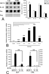
SLFs release MCP-1, but not Il-1β, upon exposure to NTHI lysate. (A) The protein array and its densitinogram show that the spiral ligament cells release MCP-1 after treatment with a lysate of NTHI, compared with the PBS-treated control. Cell culture medium was collected 48 h after treatment, and a solid-phase multiplexed protein assay in a sandwich ELISA format was performed. The chemiluminescence signal was detected by exposure to X-ray film and quantitated using Quantity One software. MCP-1 is released from SLFs after treatment with a lysate of NTHI, while release of IL-1β is not obvious. Con, PBS-treated control; Pos, positive signal produced by blotting of biotin-conjugated IgG; Neg, negative signal produced by blotting of vehicle. (B) Real-time quantitative PCR demonstrates a time-dependent up-regulation in the levels of MCP-1 mRNA after treatment with NTHI lysate, reaching saturation at between 4 h and 8 h. (C) MCP-1 expression levels are much higher than IL-1β levels in both the spiral ligament cell line and primary epidermal fibroblasts (CRL). For real-time quantitative PCR, multiplex PCR was performed and the CT values of MCP-1 were normalized to the internal control, GAPDH. Results are expressed relative to the fold induction of mRNA levels, taking the value of the nontreated group as 1. The experiments were performed in triplicate and repeated more than twice. Values are given as means ± standard deviations (n = 3). *, P < 0.05; **, P < 0.01.
FIG. 3.
FIG. 3.
TLR2 and MyD88 are involved in NTHI-induced MCP-1 up-regulation of SLFs. (A) Both conventional RT-PCR and real-time quantitative PCR show that the spiral ligament cell line expresses TLR2 and TLR4. TLR2 is up-regulated only by NTHI treatment in SLFs. Con, control. (B) NTHI-induced MCP-1 up-regulation is inhibited by the dominant-negative TLR2 construct (TLR2_DN) but not by the TLR4 dominant-negative construct (TLR4_DN) (left panel). In contrast, overexpression of wild-type TLR2 (TLR2_WT) enhances NTHI-induced MCP-1 up-regulation. The luciferase assay demonstrates that the TLR2 dominant-negative construct inhibits NTHI-induced activation of the MCP-1 promoter (right panel). MCP1-E vector was cotransfected with the TLR2 dominant-negative construct and pRL-TK vector. pcDNA, mock transfection with a blank vector. (C) Dissection microscopic views demonstrating separation of the cochlear lateral wall from the cochlea of gene-targeted mouse pups. The cochlea is isolated with preservation of its normal structure, such as the round window (arrowhead), after dissecting the inner ear from the skull base (C1). After removal of the bony otic capsule (C2), the cochlear lateral wall is separated (C3) and ready for the explant culture (C4). Apex, cochlear apex; CL, cochlear lateral wall; CoN, cochlear nerve; #, an explant of the cochlear lateral wall. (D) Real-time quantitative PCR shows that targeting TLR2 or MyD88 blocks NTHI-induced MCP-1 up-regulation more than 90% and 100%, respectively. The NTHI lysate was incubated with the primary SLFs derived from wild-type or knockout mice after starvation. Cells were harvested 3 h after treatment, and total RNA was extracted. After synthesizing cDNA, multiplex PCR was performed for real-time quantitation, and the CT values of MCP-1 were normalized to the internal control, GAPDH. Results are expressed as fold induction of mRNA quantity or luciferase activity, taking the value of the nontreated group as 1. The experiments were performed in triplicate and repeated more than twice. Values are given as the means ± standard deviations (n = 3). *, P < 0.05; **, P < 0.01.
FIG. 4.
FIG. 4.
Activation of the IκKβ-IκBα-NF-κB signaling pathways is required for NTHI-induced MCP-1 up-regulation. (A) NF-κB is translocated from the cytoplasm to the nucleus after NTHI treatment. However, CAPE and MG132 (a cell-permeative proteasome inhibitor) block the NTHI-induced NF-κB translocation. Cells were exposed to the NTHI lysate with or without pretreatment with CAPE or MG132. Cells were immunolabeled using a polyclonal anti-p65 antibody to localize cytoplasmic or nuclear NF-κB. Con, PBS-treated control. (B) Real-time quantitative PCR shows that CAPE and MG132 inhibit NTHI-induced MCP-1 up-regulation. NTHI lysate was added to the spiral ligament cell line with or without pretreatment with CAPE or MG132. Cells were harvested after 3 h, and total RNA was extracted. Multiplex PCR was performed using specific primers and probes to MCP-1 and GAPDH for real-time quantitative PCR. (C) Upstream components of the NF-κB pathway are involved in NTHI-induced MCP-1 up-regulation. Phosphorylation assay demonstrates that IκBα and IκKβ are phosphorylated by treatment with the NTHI lysate. Real-time quantitative PCR shows that transfecting a dominant-negative construct of IκBα and IκKβ inhibits NTHI-induced MCP-1 up-regulation in a dose-dependent manner. (D) The luciferase assay demonstrates that a dominant-negative construct of IκBα and IκKβ inhibits NTHI-induced activation of the MCP-1 promoter. MCP1-E vector was cotransfected with the dominant-negative constructs and pRL-TK vector. pcDNA, mock transfection with a blank vector. Results are expressed as fold induction of mRNA levels or luciferase activity, taking the value of the nontreated group as 1. The experiments were performed in triplicate and repeated more than twice. Values are given as the means ± standard deviations (n = 3). *, P < 0.05; **, P < 0.01.
FIG. 5.
FIG. 5.
Identification of NF-κB subunits and their associated binding sites involved in NTHI-induced MCP-1 up-regulation. (A) 5′ flanking regions of rat MCP-1 were subcloned into a luciferase-expressing vector. The pMCP1-E construct contained the NF-κB-binding site 3 (NF-κB3) as well as the NF-κB-binding sites of the enhancer region (NF-κB1 and NF-κB2). In contrast, the pMCP1 construct had only NF-κB3. Cells were first transfected with pGL3-B (a blank vector without a promoter region), pMCP1, or pMCP1-E and subsequently treated with NTHI lysate, and the effects measured using a luciferase assay. The assay shows that the enhancer region containing NF-κB1 and NF-κB2 is of importance for NTHI-induced MCP-1 up-regulation. Results are expressed as fold-induction of luciferase activity, taking the value of the nontreated group as 1. (B) NF-κB-binding sites of the enhancer region are conserved in mammals, i.e., mouse, rat, and human. EMSA was performed with oligonucleotides of the rat MCP-1 enhancer region, spanning the region from −2,272 to −2,297 for NF-κB site 1 and from −2,242 to −2,266 for NF-κB site 2. (C) Nuclear extract of NTHI-treated cells binds to NF-κB site 1 but not to NF-κB site 2. Two complexes (arrows) are induced in NTHI-treated cells. Lanes 1 and 4, NTHI-treated cells; lanes 2 and 5, non-NTHI-treated cells; lanes 3 and 6, oligonucleotide only; PC, positive control (biotinylated EBNA control DNA plus EBNA extract); NC, negative control (biotinylated EBNA control DNA only). (D) ELISA-based transcription factor assay shows that the p65 subunit is activated and translocated to the nucleus by treatment with the NTHI lysate. Nuclear protein was applied to a 96-well plate coated with oligonucleotides containing the consensus sequence of a different transcription factor (p65, p50, c-Rel, c-Fos, CREB-1 or ATF2). Binding of transcription factors was inhibited with a competitor DNA sequence similar to that in the oligonucleotide-coated wells, demonstrating the binding specificity between DNA and the transcription factor. Bound transcription factors were labeled first with primary polyclonal antibodies specific for each transcription factor and then with a secondary antibody, an anti-rabbit IgG antibody conjugated with HRP. After incubation with the tetramethylbenzidine substrate, the absorbance was measured at 655 nm with a microtiter plate reader. OD, optical density. The experiments were performed in triplicate and repeated more than twice. Values are given as the means ± standard deviations (n = 3). **, P < 0.01.
FIG. 6.
FIG. 6.
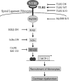
Schematic representation of the signaling pathways involved in NTHI-induced MCP-1 up-regulation. As indicated, NTHI up-regulates MCP-1 expression via TLR2-dependent NF-κB activation. It is hypothesized that the up-regulation of MCP-1 results in recruitment and attraction of effector cells, leading to cochlear dysfunction and subsequent sensorineural hearing loss.

Similar articles

Cited by

References

    1. Adachi, O., T. Kawai, K. Takeda, M. Matsumoto, H. Tsutsui, M. Sakagami, K. Nakanishi, and S. Akira. 1998. Targeted disruption of the MyD88 gene results in loss of IL-1- and IL-18-mediated function. Immunity 9:143-150. - PubMed
    1. Aderem, A., and R. J. Ulevitch. 2000. Toll-like receptors in the induction of the innate immune response. Nature 406:782-787. - PubMed
    1. Agrawal, S., M. Husein, and D. MacRae. 2005. Complications of otitis media: an evolving state. J. Otolaryngol 34(Suppl. 1):S33-S39. - PubMed
    1. Akira, S., and K. Takeda. 2004. Toll-like receptor signalling. Nat. Rev. Immunol. 4:499-511. - PubMed
    1. Akira, S., M. Yamamoto, and K. Takeda. 2003. Role of adapters in Toll-like receptor signalling. Biochem. Soc. Trans. 31:637-642. - PubMed

Publication types

MeSH terms

LinkOut - more resources