. 2007 Jul;75(7):3581-93.
doi: 10.1128/IAI.00214-07. Epub 2007 Apr 23.

Adapter protein SH2-Bbeta stimulates actin-based motility of Listeria monocytogenes in a vasodilator-stimulated phosphoprotein (VASP)-dependent fashion

Affiliations

Adapter protein SH2-Bbeta stimulates actin-based motility of Listeria monocytogenes in a vasodilator-stimulated phosphoprotein (VASP)-dependent fashion

Maria Diakonova et al. Infect Immun. 2007 Jul.

Abstract

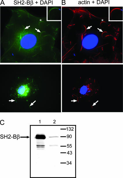
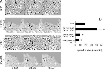
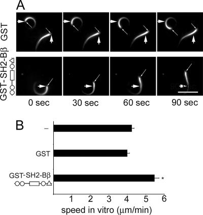
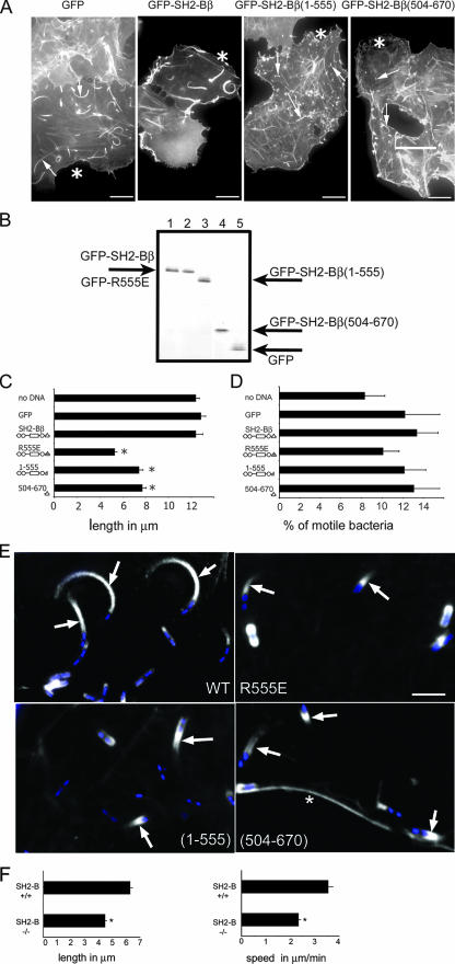
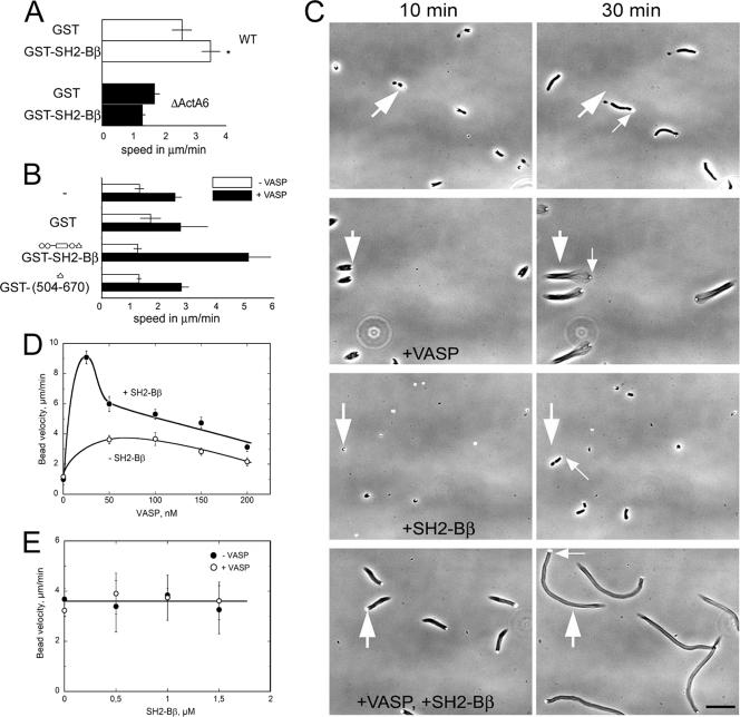
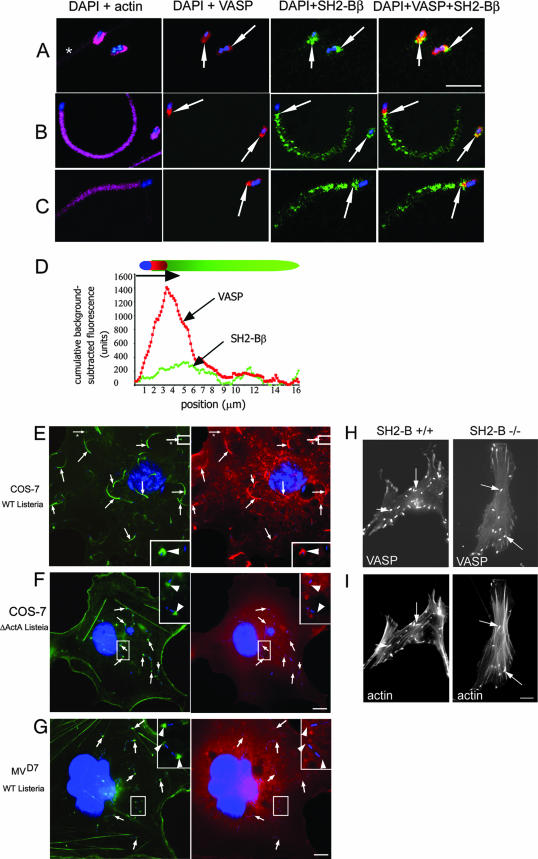
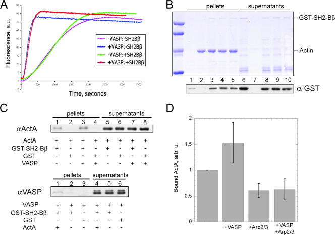
SH2-Bbeta (Src homology 2 Bbeta) is an adapter protein that is required for maximal growth hormone-dependent actin reorganization in membrane ruffling and cell motility. Here we show that SH2-Bbeta is also required for maximal actin-based motility of Listeria monocytogenes. SH2-Bbeta localizes to Listeria-induced actin tails and increases the rate of bacterial propulsion in infected cells and in cell extracts. Furthermore, Listeria motility is decreased in mouse embryo fibroblasts from SH2-B(-/-) mice. Both recruitment of SH2-Bbeta to Listeria and SH2-Bbeta stimulation of actin-based propulsion require the vasodilator-stimulated phosphoprotein (VASP), which binds ActA at the surfaces of Listeria cells and enhances bacterial actin-based motility. SH2-Bbeta enhances actin-based movement of ActA-coated beads in a biomimetic actin-based motility assay, provided that VASP is present. In vitro binding assays show that SH2-Bbeta binds ActA but not VASP; however, binding to ActA is greater in the presence of VASP. Because VASP also plays an essential regulatory role in actin-based processes in eukaryotic cells, the present results provide mechanistic insight into the functions of both SH2-Bbeta and VASP in motility and also increase our understanding of the fundamental mechanism by which Listeria spreads.


Figures

FIG. 1.
FIG. 1.
Endogenous SH2-Bβ is present in Listeria actin tails. (A) COS-7 cells were infected with Listeria and stained with anti-SH2-Bβ (green), DAPI (blue), and phalloidin-Texas Red (red). Arrows indicate bacterial actin tails. Boxed regions in the upper right corners are enlarged images of the tails marked with asterisks in the larger images. (B) Macrophages were infected with Listeria and stained with anti-SH2-Bβ (green), DAPI (blue), and phalloidin-Texas Red (red). Arrows indicate bacterial actin tails. (C) Whole-cell lysates of COS-7 cells transfected with (lane 1) or without (lane 2) cDNA encoding SH2-Bβ were subjected to anti-SH2-Bβ Western blotting. The migration of endogenous SH2-Bβ and molecular weight standards is indicated.
FIG. 2.
FIG. 2.
SH2-Bβ enhances intracellular actin-dependent Listeria motility. (A) COS-7 cells were transfected with cDNA encoding either GFP alone or the indicated forms of GFP-SH2-Bβ. The large arrows refer to the initial position of the bacterium, while small arrows indicate the moving bacterium. In the schematic of SH2-Bβ, the circles represent proline-rich regions, the rectangle represents the pleckstrin homology domain, and the triangles represent the SH2 domain. Bar, 4 μm. (B) The movement of bacteria versus time was measured, and the velocity was calculated. Bars represent means plus standard errors of the means (SEM) [n = 13, 13, 10, and 14 for cells expressing GFP, GFP-SH2-Bβ, GFP-SH2-Bβ(R555E), and GFP-SH2-Bβ(504-670), respectively]. *, P < 0.05 compared to cells expressing GFP.
FIG. 3.
FIG. 3.
SH2-Bβ enhances Listeria motility in Xenopus egg extract. (A) Listeria cells were incubated in Xenopus egg extracts supplemented with rhodamine-labeled G-actin and either GST or GST-SH2-Bβ. Large arrows indicate the initial position of the bacterium, and thin arrows indicate the moving bacterium. Bar, 8 μm. (B) The movement of bacteria versus time was measured, and the velocity was calculated. Bars represent means plus SEM (n = 8, 12, and 17 for extracts without supplementation and those supplemented with GST and GST-SH2-Bβ, respectively). *, P < 0.05 compared to extracts supplemented with GST.
FIG. 4.
FIG. 4.
SH2-Bβ affects the length of Listeria actin tails. (A) COS-7 cells expressing the indicated proteins were infected with Listeria and stained for actin. Asterisks denote transfected cells. Arrows indicate Listeria actin tails. Bars, 10 μm. (B) GFP-SH2-Bβ (lane 1), GFP-SH2-Bβ(R555E) (lane 2), GFP-SH2-Bβ(1-555) (lane 3), GFP-SH2-Bβ(504-670) (lane 4), and GFP (lane 5) were overexpressed in COS-7 cells. GFP and GFP-tagged forms of SH2-Bβ were immunoprecipitated using anti-GFP and visualized by blotting with anti-GFP. The migration of GFP and GFP-tagged forms of SH2-Bβ is indicated. (C) Lengths of actin tails of Listeria in cells expressing the indicated forms of SH2-Bβ. n = 280, 117, 98, 95, 102, and 119 for nontransfected cells and cells expressing GFP, GFP-SH2-Bβ, GFP-SH2-Bβ(R555E), GFP-SH2-Bβ(1-555), and GFP-SH2-Bβ(504-670), respectively. (D) The number of Listeria organisms with actin tails as a percentage of Listeria organisms per cell was determined for cells expressing the indicated forms of GFP-SH2-Bβ. n = 24, 15, 21, 16, 18, and 25 for nontransfected cells and cells overexpressing GFP, GFP-SH2-Bβ, GFP-SH2-Bβ(R555E), GFP-SH2-Bβ(1-555), and GFP-SH2-Bβ(504-670), respectively. (E) COS-7 cells expressing the indicated GFP-tagged proteins (white) were infected with Listeria and stained with DAPI (blue) and phalloidin-Texas Red (not shown). Arrows indicate GFP-SH2-Bβ in long bacterial tails and GFP-tagged SH2-Bβ(R555E), SH2-Bβ(1-555), and SH2-Bβ(504-670) in short Listeria tails. *, plasma membrane. Bar, 5 μm. (F) The lengths of actin tails of Listeria were determined in MEF from SH2-B+/+ and SH2-B−/− mice. n = 245 for +/+ MEF and 344 for −/− MEF. The movement of bacteria in MEF from SH2-B+/+ (n = 123) and SH2-B−/− (n = 179) mice versus time was measured, and velocities were calculated. For panels C, D, and F, bars represent means plus SEM for three independent experiments. *, P < 0.05 compared to cells expressing GFP (B) or to MEF from SH2-B+/+ mice (F).
FIG. 5.
FIG. 5.
VASP is required for the effect of SH2-Bβ on actin-based motility. (A) WT or ΔActA6 Listeria cells were incubated in Xenopus egg extract supplemented with either GST or GST-SH2-Bβ. The movement of individual bacteria was measured, and velocities were calculated. Bars represent means plus SEM. *, P < 0.05 compared to extracts supplemented with GST. (B) ActA-coated beads were incubated in motility medium, with or without 150 nM VASP and 1 μM of the indicated proteins. For each assay, the movement of n beads was measured, and the average velocity was calculated. From top to bottom, n = 5, 6, 6, and 4 without VASP and n = 7, 7, 8, and 8 with VASP. Bars represent means plus standard deviations (SD). (C) Time-lapse images of ActA-coated bead movement in motility medium, with or without 150 nM VASP and/or 1 μM SH2-Bβ, as indicated. The large arrows refer to the initial position of the bead, while small arrows indicate the moving bead. Flows in the samples sometimes induced a drift of the objects. Bar, 40 μm. (D) The velocity of ActA-coated beads was determined in motility medium supplemented with increasing concentrations of VASP in the presence (solid circles) or absence (open circles) of 1 μM SH2-Bβ. Bars represent means ± SD, calculated for a set of n beads in each sample. For increasing concentrations of VASP, n = 7, 5, 8, 6, 8, and 11 with SH2-Bβ and n = 5, 5, 9, 8, and 10 without SH2-Bβ. (E) The velocity of N-WASP-coated beads was determined in motility medium supplemented with increasing concentrations of SH2-Bβ in the presence (open circles) or absence (solid circles) of 150 nM VASP. The bead movement was independent of VASP and SH2-Bβ. Bars represent means ± SD, calculated for a set of n beads. For increasing concentrations of SH2-Bβ, n = 10, 13, 14, and 13 without VASP and n = 9, 6, 5, and 7 with VASP.
FIG. 6.
FIG. 6.
SH2-Bβ colocalizes with VASP and requires VASP for localization with actin. (A to C) COS-7 cells were infected with Listeria, stained with anti-SH2-Bβ (green), anti-VASP (red), phalloidin-Texas Red (pink), and DAPI (blue), and imaged by confocal microscopy. VASP colocalizes with SH2-Bβ on the side of the bacterium (A and B, yellow areas and arrows) and the beginning of the tail (B and C, yellow areas and arrows). *, the plasma membrane. Bar, 5 μm. (D) Cumulative fluorescence intensities for VASP (red) and SH2-Bβ (green) in the long actin tails were background subtracted and plotted as a function of position. (E and F) COS-7 cells overexpressing myc-SH2-Bβ were infected with either a WT (E) or ΔActA6 strain (F) of Listeria. Actin tails and actin caps at one bacterial pole were visualized by phalloidin-Alexa Fluor 488 (green, arrows), and myc-SH2-Bβ was visualized with an anti-myc antibody (red). Arrows from the images stained for actin and bacteria were superimposed on the images stained for myc-SH2-Bβ and bacteria and demonstrate that myc-SH2-Bβ localizes in the long actin tails and the actin caps formed by WT but not ΔActA Listeria. The boxed regions in the right corners are enlarged images of the marked areas in the larger images. Arrowheads from the images stained for actin and bacteria were superimposed on the images stained for SH2-Bβ and bacteria. Arrows with asterisks indicate actin caps formed by WT Listeria. Bar, 10 μm. (G) MVD7 fibroblasts were infected with WT Listeria. Actin caps were visualized by phalloidin-Alexa Fluor 488 (green, arrows), and myc-SH2-Bβ was visualized with anti-myc antibody (red). Arrows were superimposed as described above to demonstrate that there is no colocalization of these two proteins. The boxed regions in the upper right corners are enlarged images of the marked areas in the larger images. Arrowheads from the images stained for actin and bacteria were superimposed on the images stained for myc-SH2-Bβ and bacteria. Bar, 10 μm. (H and I) MEF from SH2-B+/+ (D) and SH2-B−/− (E) mice overexpressing GFP-VASP were infected with WT Listeria and stained for F-actin. GFP-VASP was localized at the poles of bacteria in both cell types. Arrows indicate actin tails (lower images) and GFP-VASP (upper images). Bar, 10 μm.
FIG. 7.
FIG. 7.
Interaction of SH2-Bβ with actin and ActA. (A) Actin (3 μM; 10% pyrenyl labeled) was polymerized in the presence of ActA-coated beads and the Arp2/3 complex (20 nM), with or without VASP (107 nM) and with or without SH2-Bβ (0.5 μM). Polymerization was monitored by the increase in fluorescence intensity of pyrenyl-actin. a.u., arbitrary units. (B) Actin was polymerized in the presence of SH2-Bβ and additional proteins. F-actin and G-actin were separated by high-speed centrifugation, and the proteins present in pellets (lanes 1 to 5) and supernatants (lanes 6 to 10) were detected by SDS-PAGE (top). Additional bands in the supernatants (lanes 6 and 8 to 10) are degraded forms of SH2-Bβ that do not bind F-actin, confirming that specific binding is observed in lanes 3 to 5. Immunoblotting using anti-GST antibody was done to detect SH2-Bβ with a higher contrast (bottom). Actin was polymerized alone (lanes 2 and 7), in the presence of 0.5 μM SH2-Bβ (lanes 3 and 8), or supplemented with 50 nM Arp2/3 complex and 40 nM ActA, without (lanes 4 and 9) or with (lanes 5 and 10) 50 nM VASP. The control without actin (lanes 1 and 6) shows a slight contamination (<5%) of the pellet by SH2-Bβ that is negligible compared to the amount of SH2-Bβ (∼30%) recruited by F-actin. (C) Pull-down assays of ActA and VASP with SH2-Bβ. (Top) ActA binds to GST-SH2-Bβ in the absence (lane 1) and, more strongly, in the presence (lane 3) of VASP. No ActA was detected in the negative controls (GST alone) (lanes 2 and 4). (Bottom) VASP alone (lane 2) does not bind to GST-SH2-Bβ beads and is recruited only via its strong interaction with ActA (lane 1). (D) Binding of ActA to SH2-Bβ as a function of Arp2/3 and VASP. In each experiment, the amount of ActA bound to GST-SH2-Bβ in the absence of other ligands is taken as a reference and normalized to 1 (left bar). The addition of VASP increased the amount of bound ActA by 50%. The addition of Arp2/3 induced a decrease of ∼40%, with or without VASP. Bars represent means plus SD, computed from three independent experiments.

Similar articles

Cited by

References

    1. Barzik, M., T. I. Kotova, H. N. Higgs, L. Hazelwood, D. Hanein, F. B. Gertler, and D. A. Schafer. 2005. Ena/VASP proteins enhance actin polymerization in the presence of barbed end capping proteins. J. Biol. Chem. 280:28653-28662. - PMC - PubMed
    1. Bear, J. E., J. J. Loureiro, I. Libova, R. Fassler, J. Wehland, and F. B. Gertler. 2000. Negative regulation of fibroblast motility by Ena/VASP proteins. Cell 101:717-728. - PubMed
    1. Bear, J. E., T. M. Svitkina, M. Krause, D. A. Schafer, J. J. Loureiro, G. A. Strasser, I. V. Maly, O. Y. Chaga, J. A. Cooper, G. G. Borisy, and F. B. Gertler. 2002. Antagonism between Ena/VASP proteins and actin filament capping regulates fibroblast motility. Cell 109:509-521. - PubMed
    1. Boujemaa-Paterski, R., E. Gouin, G. Hansen, S. Samarin, C. Le Clainche, D. Didry, P. Dehoux, P. Cossart, C. Kocks, M. F. Carlier, and D. Pantaloni. 2001. Listeria protein ActA mimics WASp family proteins: it activates filament barbed end branching by Arp2/3 complex. Biochemistry 40:11390-11404. - PubMed
    1. Cicchetti, G., P. Maurer, P. Wagener, and C. Kocks. 1999. Actin and phosphoinositide binding by the ActA protein of the bacterial pathogen Listeria monocytogenes. J. Biol. Chem. 274:33616-33626. - PubMed

Publication types

MeSH terms

LinkOut - more resources