. 2007 Jul;51(7):2642-5.
doi: 10.1128/AAC.00007-07. Epub 2007 Apr 23.

Sultam thiourea inhibition of West Nile virus

Affiliations

Sultam thiourea inhibition of West Nile virus

Eric Barklis et al. Antimicrob Agents Chemother. 2007 Jul.

Abstract

We have identified sultam thioureas as novel inhibitors of West Nile virus (WNV) replication. One such compound inhibited WNV, with a 50% effective concentration of 0.7 microM, and reduced reporter expression from cells that harbored a WNV-based replicon. Our results demonstrate that sultam thioureas can block a postentry, preassembly step of WNV replication.


Figures

FIG. 1.
FIG. 1.
Compound structures. The diagrams show the structures of the following sultams: TYT-1, N′-(1,1-dioxido-2-phenyl-1,4,2-dithiazolidin-3- ylidene)-N,N-diphenylthiourea (439.6 kDa); TYT-2, (2,5-dimethyl-1,1-dioxido-1,4,2-dithiazolidin-3-ylidene)bis(1-methylethyl)thiourea (323.5 kDa); TYT-3, [1,1-dioxido-2-(phenylmethyl)-1,4,2-dithiazolidin-3-ylidene]bis(1- methylethyl)-thiourea (385.6 kDa); and TYT-4, (1,1-dioxido-2-phenyl-1,4,2-dithiazolidin-3-ylidene)-bis(phenylmethyl)-thiourea (467.6 kDa).
FIG. 2.
FIG. 2.
WNV yield reduction. Vero cells in medium containing 100 U/ml penicillin and 0.1 mg/ml streptomycin were mock treated with DMSO (no TYT-1; no virus; final DMSO concentration, 1%) or treated with the indicated concentration of TYT-1 in DMSO and then mock infected (no virus) or infected with WNV at an MOI of 1.0. At 24 h p.i., virus-containing medium samples at the indicated dilutions were used to infect fresh cells. At 5 days p.i., surviving cells were stained with 0.0375% crystal violet. Infected, mock-treated wells were devoid of cells due to WNV-mediated cell killing, and wells were routinely scored as virus positive if cell staining levels were reduced three-fourths or more relative to uninfected cell staining levels. Note that 2.3 and 23 μM TYT-1 reduced WNV titers at least 100-fold and that these results are representative of more than 10 independent experiments.
FIG. 3.
FIG. 3.
Effective anti-WNV drug concentrations. Increasing concentrations of TYT-1 (black bars), TYT-2 (striped bars), TYT-3 (gray bars), and TYT-4 (white bars) were used to determine effective anti-WNV concentrations by virus yield reduction assays. Results are plotted as concentrations versus percentages of virus yields observed in mock-treated (1% DMSO [final concentration]) controls. Titers were determined by limiting dilution and scored as described in the legend to Fig. 2. Averages (means) were derived from three separate experiments for TYT-1 and at least two separate experiments for TYT-2 to -4, and standard deviations are shown.
FIG. 4.
FIG. 4.
WNV RNA levels in treated and untreated cells. Vero cells were mock treated with DMSO (“mock”; final concentration, 0.5% DMSO) or treated with 11 μM TYT-1 in DMSO (“TYT-1”) and then mock infected (not shown) or infected with WNV at an MOI of 5. At 18 h p.i., RNAs were isolated, and equivalent input RNA amounts were reverse transcribed and subjected to real-time PCR quantitation of WNV RNA levels following previously described protocols (8, 19). The results, plotted as relative fluorescence signals versus PCR cycle numbers, indicate the following average (n = 4) WNV RNA copy numbers per cell, as quantitated relative to an in vitro-transcribed NS3 RNA standard: for uninfected cells, 0; for untreated cells, 3,255 ± 325.8; and for TYT-1-treated cells, 27.2 ± 1.6. Reverse transcription-PCR cycle parameters were 30 min at 48°C for the reverse transcription step, 10 min at 95°C for a denaturation step, and 40 cycles of 13 s at 95°C and 1 min at 60°C.
FIG. 5.
FIG. 5.
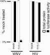
WNV replicon inhibition. BHK cells expressing a WNV luciferase replicon (WNV) (33) or 293 cells transfected with an HIV-based luciferase expression vector (HIV) (34) were mock treated with DMSO (0.1% [final concentration]) or treated with 23 μM TYT-1 in DMSO. At 48 h posttreatment, cells were processed for detection of total protein levels (white bars) or luciferase activities (black bars). Protein levels and luciferase levels are expressed as percentages of the values obtained for mock-treated samples; background luciferase levels with parental BHK and untransfected 293 cells were <0.1% of the 100% values shown. Values obtained for WNV replicon samples were averaged from four separate experiments and are shown with standard deviations.

Similar articles

Cited by

References

    1. Abbenante, G., and D. Fairlie. 2005. Protease inhibitors in the clinic. Med. Chem. 171-104. - PubMed
    1. Alfadhli, A., Z. Love, B. Arvidson, J. Seeds, J. Willey, and E. Barklis. 2001. Hantavirus nucleocapsid protein oligomerization. J. Virol. 752019-2023. - PMC - PubMed
    1. Anonymous. 2003. Tricyclic lactam and sultam derivatives as histone deacetylase inhibitors. Expert Opin. Ther. Pat. 13387-391.
    1. Arvidson, B., J. Seeds, M. Webb, L. Finlay, and E. Barklis. 2003. Analysis of the retrovirus capsid interdomain linker region. Virology 308166-177. - PubMed
    1. Baker, D. C., and B. Jiang. March 2002. Sultams: solid phase and other synthesis of anti-HIV compounds and compositions. U.S. patent 6,353,112 B1.

Publication types

MeSH terms

LinkOut - more resources