. 2007 May 1;104(18):7682-7.
doi: 10.1073/pnas.0611448104. Epub 2007 Apr 23.

A monomeric G protein-coupled receptor isolated in a high-density lipoprotein particle efficiently activates its G protein

Affiliations

A monomeric G protein-coupled receptor isolated in a high-density lipoprotein particle efficiently activates its G protein

Matthew R Whorton et al. Proc Natl Acad Sci U S A. .

Abstract

G protein-coupled receptors (GPCRs) respond to a diverse array of ligands, mediating cellular responses to hormones and neurotransmitters, as well as the senses of smell and taste. The structures of the GPCR rhodopsin and several G proteins have been determined by x-ray crystallography, yet the organization of the signaling complex between GPCRs and G proteins is poorly understood. The observations that some GPCRs are obligate heterodimers, and that many GPCRs form both homo- and heterodimers, has led to speculation that GPCR dimers may be required for efficient activation of G proteins. However, technical limitations have precluded a definitive analysis of G protein coupling to monomeric GPCRs in a biochemically defined and membrane-bound system. Here we demonstrate that a prototypical GPCR, the beta2-adrenergic receptor (beta2AR), can be incorporated into a reconstituted high-density lipoprotein (rHDL) phospholipid bilayer particle together with the stimulatory heterotrimeric G protein, Gs. Single-molecule fluorescence imaging and FRET analysis demonstrate that a single beta2AR is incorporated per rHDL particle. The monomeric beta2AR efficiently activates Gs and displays GTP-sensitive allosteric ligand-binding properties. These data suggest that a monomeric receptor in a lipid bilayer is the minimal functional unit necessary for signaling, and that the cooperativity of agonist binding is due to G protein association with a receptor monomer and not receptor oligomerization.


Conflict of interest statement

The authors declare no conflict of interest.

Figures
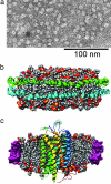
Fig. 1.
Fig. 1.
Depiction of rHDL particles. (a) Transmission electron micrograph of negatively stained rHDL. Well defined 10-nm rHDL particles are clearly visible. (Scale bar, 100 nm.) (b) Molecular model illustrating rHDL composed of a dimer of apoA-I proteins wrapped around a phospholipid bilayer composed of 160 POPC molecules (24). Each apoA-I protein (cyan and green) is depicted as a ribbon diagram. (c) Molecular model of a GPCR (bovine rhodopsin, Protein Data Bank ID code 1F88) reconstituted into rHDL. Images were generated by using UCSF Chimera Package from the Resource for Biocomputing, Visualization, and Informatics at the University of California, San Francisco (supported by National Institutes of Health Grant P41 RR-01081) (44). Coordinates for the HDL model, from Segrest et al. (24), are used with permission from Stephen Harvey, Georgia Institute of Technology, Atlanta, GA.
Fig. 2.
Fig. 2.
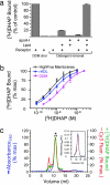
Functional reconstitution of β2AR into rHDL. (a) β2AR requires apoA-I and lipids to survive detergent removal with high efficiency. Equal amounts of DDM-solubilized β2AR were included in the rHDL reconstitution assay with or without apoA-I and lipid (POPC/palmitoyl-oleoyl-phosphatidylglycerol at 3:2) as described in Materials and Methods and assayed for [3H]DHAP binding (at 20 nM). For comparison, equal amounts of receptor were also assessed for binding activity in the presence of 25 mM cholate (“cho”) or 1% DDM. Data shown are specific binding. (b) Saturation-binding assays were performed on β2AR in HighFive cell membranes (open squares), in DDM micelles (pink filled circles), or in rHDL lipid bilayers (blue filled squares). (c) Cy3-β2AR·rHDL elutes as a single peak by SEC (Superdex 200; GE Healthcare) as monitored by UV absorbance (blue), [3H]DHAP binding (green), and fluorescence (red). Bracketed area (with asterisk) represents the fractions analyzed for functional and fluorescence studies. (Inset) Preparations of Cy5-β2AR·rHDL (red) or (Cy3-β2AR+Cy5-β2AR)·rHDL (black) elute similarly Cy3-β2AR·rHDL (blue), with a Stokes diameter of ≈11 nm.
Fig. 3.
Fig. 3.
TIRF of Cy3- and Cy5-labeled β2AR in rHDL reveals that the vast majority of β2AR are monomeric. Conditions for the reconstitutions illustrated are: (a) Cy3-β2AR·rHDL and (b) Cy5-β2AR·rHDL particles alone, or (c) Cy3-β2AR·rHDL and Cy5-β2AR·rHDL mixed together at equal concentrations, (d) Cy3-β2AR and Cy5-β2AR mixed together at equal concentrations before reconstitution (Cy3-β2R+Cy5-β2R)·rHDL, (e) β2AR colabeled with Cy3 and Cy5 in 0.1% DDM, and (f) bar graph summarizing TIRF data in c–e. Cy3- and/or Cy5-labeled receptors were reconstituted in rHDL at a β2AR:ApoA-I ratio of 1:100 (β2AR:rHDL ratio of 1:50) in the presence of POPC/POPG and resolved by SEC. Receptor concentrations were maintained at 1 μM. Fractions were analyzed by TIRF as described in Materials and Methods.
Fig. 4.
Fig. 4.
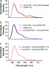
FRET measurements confirm monomeric β2AR in rHDL. Cy3- and Cy5-labeled β2AR preparations were reconstituted under different conditions, and normalized spectra were analyzed for the presence of FRET, as indicated by increased acceptor (Cy5) emission (at 670 nm) and the concomitant decrease in donor (Cy3) emission (at 575 nm). (a) Equal amounts of Cy3-β2AR and Cy5-β2AR were mixed and reconstituted into phospholipid vesicles (rVesicles). (Cy3-β2AR + Cy5-β2AR)·rVesicles display FRET, which is reduced on exposure to 1% DDM (red), a detergent concentration that disrupts and resolubilizes the vesicles. (b) In contrast, no significant FRET is observed in (Cy3-β2AR + Cy5-β2AR)·rHDL in the absence (black) or presence (red) of 1% DDM. These spectra are identical to spectra obtained from equal mixtures of Cy3-β2AR·rHDL and Cy5-β2AR·rHDL (blue). (c) Normalized spectra are also identical to mixtures of Cy3-β2AR and Cy5-β2AR in DDM micelles at 0.1% DDM (black) or at 1% (red).
Fig. 5.
Fig. 5.
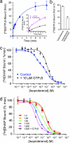
Monomeric β2AR incorporated in rHDL particles couples efficiently to G proteins. (a) ISO-induced [35S]GTPγS binding to monomeric-β2AR·Gs·rHDL particles. β2AR·Gs·rHDL particles were preincubated with either 1 μM ISO (magenta) or 10 μM timolol (black) in the presence of 100 nM [35S]GTPγS and 2 mM MgCl2 to stimulate [35S]GTPγS binding (Inset). Aliquots (at times indicated) were removed, and binding was terminated with a quench buffer. Agonist-specific [35S]GTPγS binding to β2AR·Gs·rHDL particles (blue) represents the difference between ISO-stimulated and timolol-bound data (Inset). (b) Propranolol inhibition of ISO-stimulated [35S]GTPγS binding. β2AR·Gs·rHDL particles were preincubated with either 1 μM ISO or 1 μM ISO plus 3 mM propranolol and then assayed for [35S]GTPγS (2 min at 30°C), as above. (c) High-affinity ISO inhibition of [3H]DHAP binding to monomeric β2AR in rHDL occurs in the presence of G proteins (blue) but is abolished by coincubation of 10 μM GTPγS (black). The Khigh (8.0 ± 2.4 nM) and Klow (360 ± 110 nM) sites represent 56% and 44% of total binding, respectively. Purified Gs was added to preformed β2AR·rHDL particles at a final R:G ratio of 1:0.53, as determined by [35S]GTPγS binding and described in Materials and Methods. (d) Increasing the concentration of Gs and thus the R:G ratio (e.g., R:G of 1:200) increases the proportion of high-affinity agonist sites to >90%. Note that the 1:200 represents the initial R:G ratio and not the final functional R:G (see text). Coincubation with 10 μM GTPγS to the 1:200 reconstitution condition completely abolishes the high-affinity ISO binding (open circles), yielding a Ki similar to the Klow. These data were fit with a nonlinear regression (in c and d) or two-phase exponential fit using Prism 4.0 (in a) (GraphPad).

References

    1. Catterall WA. Annu Rev Biochem. 1995;64:493–531. - PubMed
    1. Clapham DE, Runnels LW, Strubing C. Nat Rev Neurosci. 2001;2:387–396. - PubMed
    1. Dingledine R, Borges K, Bowie D, Traynelis SF. Pharmacol Rev. 1999;51:7–61. - PubMed
    1. Schlessinger J. Cell. 2000;103:211–225. - PubMed
    1. Chabre M, le Maire M. Biochemistry. 2005;44:9395–9403. - PubMed

Publication types

MeSH terms