Clinical Trial
. 2007 Apr-May;14(3):169-80.
doi: 10.1080/10739680601139179.

Noninvasive measure of microvascular nitric oxide function in humans using very low-frequency cutaneous laser Doppler flow spectra

Affiliations
Clinical Trial

Noninvasive measure of microvascular nitric oxide function in humans using very low-frequency cutaneous laser Doppler flow spectra

Julian M Stewart et al. Microcirculation. 2007 Apr-May.

Abstract

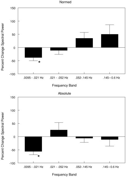
Objective: While higher frequency oscillations (0.021-0.6 Hz) in cutaneous blood flow measured by laser Doppler flowmetry (LDF) relate to oscillations in blood pressure and sympathetic nerve activity, very low-frequency oscillations (VLF, 0.0095-0.021 Hz) do not. The authors investigated whether VLF LDF power is nitric oxide (NO) specific.

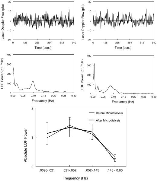
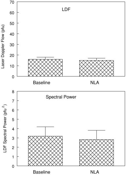
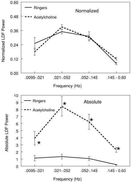
Methods: LDF combined with intradermal microdialysis was used in the calves of 22 healthy volunteers aged 19-27 years. LDF power spectral analysis was performed by windowed fast Fourier transform. The authors tested whether the NO synthesis inhibitor nitro-l-arginine (NLA) produced selective decreases in VLF power before and after stimulation with acetylcholine.

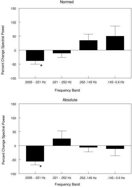
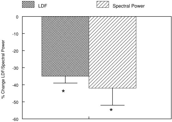
Results: NLA alone did not alter total power but selectively reduced VLF power by approximately 50%. LDF and spectral power increased markedly across all spectra with acetylcholine. This increase was blunted by NLA, which selectively reduced VLF power by approximately 50%.

Conclusions: The data suggest that VLF oscillations in the laser Doppler signal are NO dependent, increase with cholinergic stimulation, and have potential as a noninvasive marker for NO-dependent microvascular reactivity.


Figures

Figure 1
Figure 1
(Top) Laser Doppler flow (LDF) and corresponding power spectrum before (left) and after (right) insertion of the microdialysis catheters. Patients recovered from the insertion procedure for at least 1 h. (Bottom) The absolute summed laser Doppler power in each frequency band. There is no significant difference before or after recovery from microdialysis probe insertion. VLF = 0.0095–0.021 Hz, LF = 0.021–0.052 Hz, MF = 0.052–0.145 Hz, HF = 0.145–0.6 Hz.
Figure 2
Figure 2
The effects of NLA on absolute laser Doppler blood flows (LDF), expressed in perfusion units (pfu in the upper panels), and total spectral power summed over all frequency bands, (pfu2 in the lower panels). Comparison is between the baseline determined after the microdialysis probe has been inserted and after NLA is given. No differences in laser Doppler flow or total power are evident.
Figure 3
Figure 3
Representative LDF and corresponding power spectra: (Left) Results during perfusion with Ringer’s solution; (right) results during perfusion with 10 mM NLA dissolved in Ringer’s solution. There is marked decrease in very low-frequency power. VLF = 0.0095–0.021 Hz, LF = 0.021–0.052 Hz, MF = 0.052–0.145 Hz, HF = 0.145–0.6 Hz.
Figure 4
Figure 4
The effects of NLA on spectral power at each frequency band. Percent changes in spectral power from baseline are shown. (Top) Changes in data normalized to total power. (Bottom) Changes in absolute spectral power. Very low-frequency power is only and exclusively reduced following the addition of NLA. * p < .05 compared to baseline. VLF = 0.0095–0.021 Hz, LF = 0.021–0.052 Hz, MF = 0.052–0.145 Hz, HF = 0.145–0.6 Hz.
Figure 5
Figure 5
The effects of acetylcholine (100 mM) on laser Doppler flow in all subjects (N = 22). (Top) Data normalized to total power (the sum of the power in all frequency bands). (Bottom) The absolute spectral power in units of pfu2. Acetylcholine markedly increases absolute power compared to baseline (Ringer solution) but normalized power is unaffected. This implies that power in each frequency band is increased by the same fraction. * p < .05 compared to Ringer’s solution. VLF = 0.0095–0.021 Hz, LF = 0.021–0.052 Hz, MF = 0.052–0.145 Hz, HF = 0.145–0.6 Hz.
Figure 6
Figure 6
The effects of NLA during concurrent acetylcholine (AcCh) infusion on percent change in laser Doppler flow (LDF) and total spectral power compared to acetylcholine alone summed over all frequency bands. Addition of NLA to acetylcholine marked reduces flow and spectral power. * p < .05 compared to acetylcholine alone.
Figure 7
Figure 7
The effects of NLA on spectral power at each frequency band during concurrent acetylcholine infusion. Percent changes from acetylcholine alone are shown. (Top) Changes in data normalized to total power. (Bottom) Changes in absolute spectral power. Very low-frequency power is reduced following the addition of NLA. * p < .05 compared to acetylcholine alone. VLF = 0.0095–0.021 Hz, LF = 0.021–0.052 Hz, MF = 0.052–0.145 Hz, HF = 0.145–0.6 Hz.

Similar articles

Cited by

References

    1. Funk W, Endrich B, Messmer K, Intaglietta M. Spontaneous arteriolar vasomotion as a determinant of peripheral vascular resistance. Int J Microcirc Clin Exp. 1983;2:11–25. - PubMed
    1. Mayer S. Studien zur physiologie des herzens und der blutgefässe. V. Ueber spontane blut-druckschwankungen. Akad Wiss Wien Math Nat Kl. 1876;74:281–307.
    1. Zhang R, Iwasaki K, Zuckerman JH, Behbehani K, Crandall CG, Levine BD. Mechanism of blood pressure and R-R variability: insights from ganglion blockade in humans. J Physiol. 2002;543:337–348. - PMC - PubMed
    1. Cohen MA, Taylor JA. Short-term cardiovascular oscillations in man: measuring and modelling the physiologies. J Physiol. 2002;542:669–683. - PMC - PubMed
    1. Braverman IM, Keh A, Goldminz D. Correlation of laser Doppler wave patterns with underlying microvascular anatomy. J Invest Dermatol. 1990;95:283–286. - PubMed

Publication types