. 2007 May;5(5):e117.
doi: 10.1371/journal.pbio.0050117.

Unmasking activation of the zygotic genome using chromosomal deletions in the Drosophila embryo

Affiliations

Unmasking activation of the zygotic genome using chromosomal deletions in the Drosophila embryo

Stefano De Renzis et al. PLoS Biol. 2007 May.

Erratum in

  • PLoS Biol. 2007 Aug;5(8):e213
  • PLoS Biol. 2007 Jul;5(7):e195

Abstract

During the maternal-to-zygotic transition, a developing embryo integrates post-transcriptional regulation of maternal mRNAs with transcriptional activation of its own genome. By combining chromosomal ablation in Drosophila with microarray analysis, we characterized the basis of this integration. We show that the expression profile for at least one third of zygotically active genes is coupled to the concomitant degradation of the corresponding maternal mRNAs. The embryo uses transcription and degradation to generate localized patterns of expression, and zygotic transcription to degrade distinct classes of maternal transcripts. Although degradation does not appear to involve a simple regulatory code, the activation of the zygotic genome starts from intronless genes sharing a common cis-element. This cis-element interacts with a single protein, the Bicoid stability factor, and acts as a potent enhancer capable of timing the activity of an exogenous transactivator. We propose that this regulatory mode links morphogen gradients with temporal regulation during the maternal-to-zygotic transition.


Conflict of interest statement

Competing interests. The authors have declared that no competing interests exist.

Figures

Figure 1
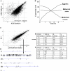
Figure 1. Time-Course Analysis of the MZT and Ablation of the Left Arm of the Second Chromosome
(A) Scatter plot representation of freshly fertilized eggs (0–1 h = 0.5 h) and mid-cycle 14 embryos (2–3 h = 2.5 h) microarray measurements. This result represents the average of two biological replicates. Note the extensive degradation of maternal mRNAs appearing as a large number of dots below the 3-fold diagonal lines. Each dot corresponds to an individual probe. The labeling “Duplicates OreR” in the x-axis of the scatter plot refer to the fly strain (Oregon R) used to collect the WT embryos. (B) Cartoon indicating the three possible outcomes of time-course microarray measurements during the MZT. (C) Scatter plot analysis of embryos missing the left arm of the second chromosomes (2L−) compared to similarly staged cycle 14 embryos mutant for the halo gene. Both population of embryos were identified based on the halo phenotype, see also Figure 2E. Results represent the average of four biological replicates. (D) Genes whose expression was down-regulated 3-fold in the 2L− collection map predominately to 2L. (E and F) Chromosomal distribution of down-regulated (E) and up-regulated (F) genes at different cut-off (p < 0.001). Only down-regulated genes were enriched on 2L.
Figure 2
Figure 2. Ablation of the X, 2-Entire, 3-Entire Chromosomes and Identification of Mutant Embryos
(A, C, E, and H) Live embryos were imaged using an up-right microscope. Arrowhead indicates the phenotypes associated with each chromosomal ablation. (A) shows a WT embryo. In (C), an X− embryo shows the irregular cellularization front (nullo phenotype) due to the failure to form furrow canals around some nuclei. (E) The 2− embryo developed the characteristic halo phenotype: a dark cytoplasmic halo below the nuclei. (H) The 3− embryo developed the bottleneck phenotype. Nuclei failed to be incorporated in the cellularization front because of the early and uncontrolled contractility of the actin-myosin network. (B, D, F, G, I, and J) Immunostaining using anti-Armadillo (B and D) and anti Myosin-2 antibodies (F, G, I, and J). (B) and (D) show the apical surface, top view, of WT and X− embryos respectively. Arrowhead indicates the irregular conformation of the apical membrane in the X− embryo. (F and G) Optical cross section of WT (F) and 2− (G) embryos stained with anti-myosin-2 antibodies. Note the lack of cell membranes in 2− embryos and failure to localize myosin-2 to the basal side of the cellularization front. (I and J) Top view of WT (I) and 3− (J) embryos stained with anti-myosyn-2 antibodies showing the typical bottleneck phenotype, arrowhead in (J). (K) Table summarizing the results of each chromosomal manipulation. The chromosomal location of down-regulated genes (3-fold, p < 0.001) is plotted next to the corresponding embryo. Data were obtained using four biological replicates (ablation of chromosome X and 2) and two biological replicates (ablation of Chromosome 3 entire).
Figure 3
Figure 3. Motif Analysis of Maternal and Zygotic Genes
(A) The complete sets of zygotic genes identified in the chromosomal deletion experiments (using the 3-fold cut-off and p < 0.001) were plotted in the time-course experiments (two biological replicates for each time point. The scatter plot was generated using the average of the two measurements). This graph demonstrates that zygotic genes can be up-regulated, down-regulated, or remain unchanged during the MZT. Increase in gene expression over time can only identify one third of the zygotic genes. (B) Stability of maternal transcripts: the expression value of 2L genes in freshly fertilized eggs (0.5 h, the average of two biological replicates is plotted) was compared to the expression value of these genes in embryos missing 2L at (2.5 h, average of four biological replicates is plotted). Under this condition, the stability of a specific transcript is solely dependent on its decay kinetics. (C) Motif discovery in the 3′ UTR of maternal unstable transcripts (upper table, the first motif is in red and the second is in blue) and in the 2-kb upstream regions of purely zygotic genes (lower table, in green). The UUGUU sequence resembles the target site for the PUF family of RNA-binding proteins, and the UAUUUAU motif resembles the AU-rich element. (D) BDGP in situ database analysis showing the expression pattern of the different categories of zygotic genes. Stage 1–3 corresponds to maternal stages (0–2 h), and 4–6 to zygotic stages (2–4 h).
Figure 4
Figure 4. The Activation of the Zygotic Genome Starts from Intronless Genes Sharing the CAGGTAG Motif
(A) Expression profile of freshly fertilized eggs (0.5 h, average of two biological replicates) compared to 1.5-h embryos (cycle 10–12, post-pole cell formation; average of two biological replicates). A group of 59 genes is significantly up-regulated at 1.5 h. Intronless genes are shown in black. About 70% of the up-regulated genes do not contain introns. (B and C) Identification of the CAGGTAG motif within the 2-kb upstream regions of the 59 genes up-regulated in (A). (C) Distribution of distances between CAGGTAG occurrences and the transcription start site of the early zygotic genes.
Figure 5
Figure 5. Purification of BSF as the 7mer Interacting Protein
(A) Coomassie staining of 7mer interacting protein. Forty grams of embryo extract was prepared as described in Materials and Methods, and subjected to two sequential purification steps. In the first step, proteins were loaded onto an agarose column coupled to the UAS. Unbound proteins were then split into two equal fractions, and each fraction was loaded onto the 7mer or UAS columns. Bound proteins were eluted with a step concentration of KCl and resolved on SDS-Page. Gel shows the results of this last purification step. One major interacting protein bound specifically to the 7mer oligo. Mass spectroscopy sequencing identified this protein as the BSF. (B) BSF binds directly and specifically to the 7mer. BSF was in vitro transcribed and translated in the presence of 35S-methionine (35S-BSF; upper panel), and the binding to the 7mer or to a mutated sequence (MUT; the two GG at position 3 and 4 were mutated to TT) was tested (lower panel). (C–E) Confocal microscopy analysis of BSF localization in the early embryo. A BSF polyclonal antibody was used to detect the endogenous localization of BSF. BSF was localized to both the cytoplasm and nuclei (arrowheads in [C] and [D]) of the blastoderm epithelium. In pole cells, BSF was localized to cytoplasmic structures, which appeared as dots, arrowhead (F). The arrow in (C) indicates the region which is enlarged in (E). The asterisk (*) in (C) indicate the region which is enlarged in (D).
Figure 6
Figure 6. The CAGGTAG Motif Activates Transcription prior to Cycle 14
(A) Transgenic embryos over-expressing GFP with (7mer) or without (control) the 7mer. Five copies of the 7mer were obtained by crossing males carrying the specific transgene to females providing Gal4. GFP expression was monitored using video microscopy. In (A), a single frame is shown. (B) Fluorescent GFP quantification at 20-min intervals. (C) FISH showing the GAL4 dependence of the 7mer-mediated GFP transcription. Only one major dot is observed per nucleus because males carrying the transgene were crossed to females supplying Gal4. (D) FISH analysis of embryos over-expressing GFP with (7mer) or without (control) the 7mer. Images were processed and quantified as described in Materials and Methods. (E and F) Quantification of the number and size of nuclear dots. Results were expressed as number of dots per 1 × 105 pixels. In (F), only dots larger than ten pixels were quantified. (G) RT-PCR quantification. Control and 7mer embryos were harvested at the indicated cycles (12 and 14), and the amount of GFP transcripts quantified using the FluorChem gel documentation system equipped with a CCD camera. The numbers below the bands are the result of this measurement. ND, non-detected.
Figure 7
Figure 7. Proposed Mode of Action for the 7mer/BSF Interaction in Timing the Response to a Morphogen Gradient
A maternally provided morphogen, for example, Dorsal, activates target gene expression in a concentration-dependent manner, defining distinct developmental units (colored rectangles). Gene a, whose enhancer has the lower affinity for this particular morphogen, is expressed only at the higher concentration of the gradient. Gene d, whose enhancer has the higher affinity, is expressed also at a lower concentration. The 7mer sequence can time the response to this gradient while maintaining the spatial information. Because the 7-mer alone is not able to activate transcription (Figure 6), it can function as a timer linking the spatial gradient with the temporal regulation. For example, the 7mer sequence contained in the 5′ regulatory region of gene a would allow gene a to be expressed before gene e within the same spatial unit (red). This regulatory mode can operate along both the dorsal-ventral (D-V) and anterior-posterior (A-P) axes because it would not interfere with the positional information encoded in the gradient itself. Indeed the 7mer and its variants are particularly enriched in enhancers bound by Dorsal (D-V) and Bicoid (A-P).

Comment in

Similar articles

Cited by

References

    1. Wieschaus E. Embryonic transcription and the control of developmental pathways. Genetics. 1996;142:5–10. - PMC - PubMed
    1. Bashirullah A, Cooperstock RL, Lipshitz HD. Spatial and temporal control of RNA stability. Proc Natl Acad Sci U S A. 2001;98:7025–7028. - PMC - PubMed
    1. Newport J, Kirschner M. A major developmental transition in early Xenopus embryos: II. Control of the onset of transcription. Cell. 1982;30:687–696. - PubMed
    1. Hamatani T, Carter MG, Sharov AA, Ko MS. Dynamics of global gene expression changes during mouse preimplantation development. Dev Cell. 2004;6:117–131. - PubMed
    1. Mathavan S, Lee SG, Mak A, Miller LD, Murthy KR, et al. Transcriptome analysis of zebrafish embryogenesis using microarrays. PLoS Genet. 2005;1:260–276. doi: 10.1371/journal.pgen.0010029. - DOI - PMC - PubMed

Publication types

LinkOut - more resources