. 2007 Aug 1;405(3):559-68.
doi: 10.1042/BJ20061653.

Mechanism of short-term ERK activation by electromagnetic fields at mobile phone frequencies

Affiliations

Mechanism of short-term ERK activation by electromagnetic fields at mobile phone frequencies

Joseph Friedman et al. Biochem J. .

Retraction in

Expression of concern in

Abstract

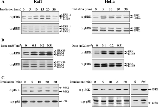
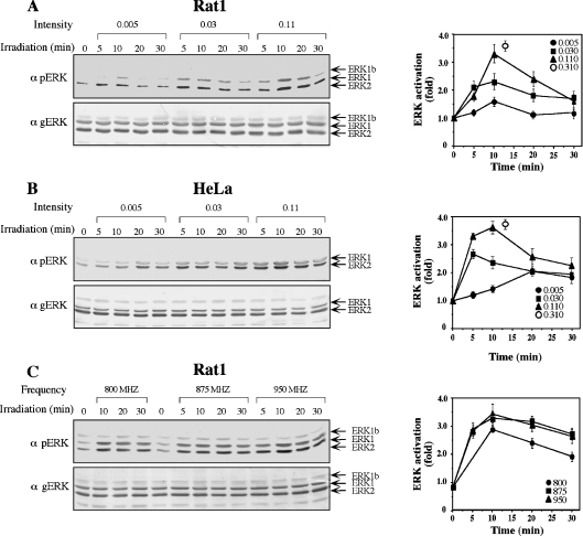
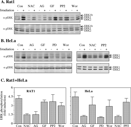
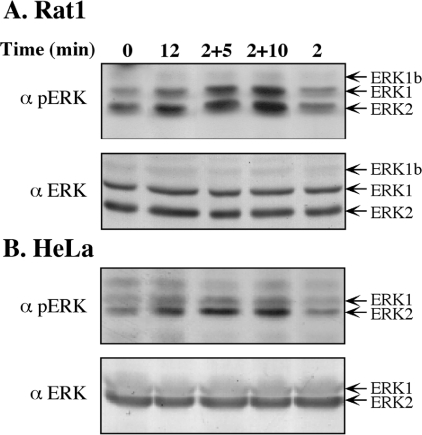
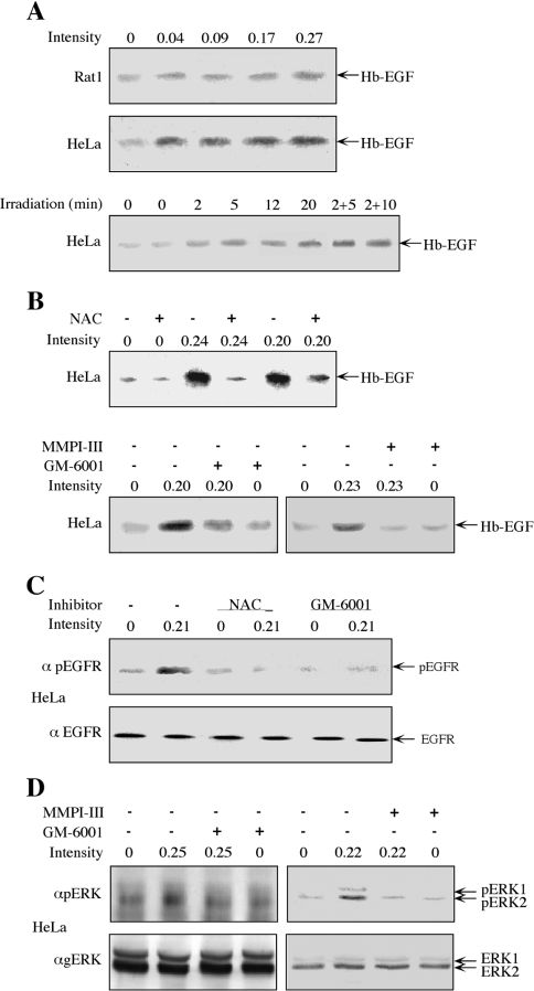
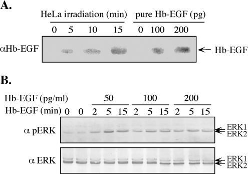
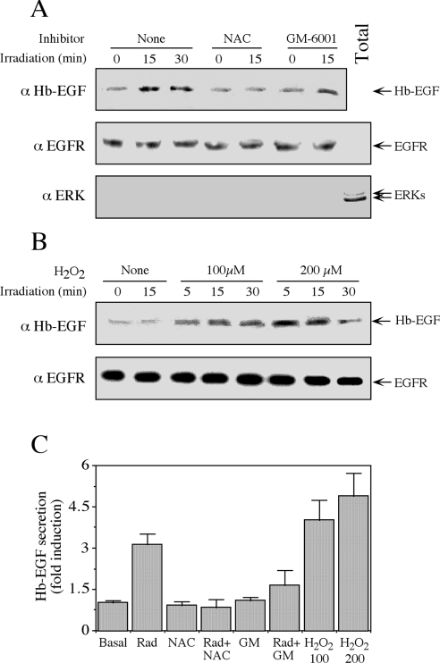
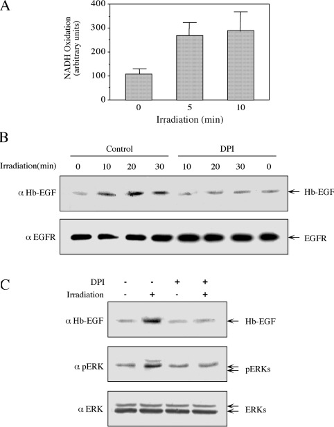
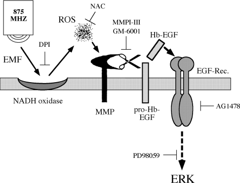
The exposure to non-thermal microwave electromagnetic fields generated by mobile phones affects the expression of many proteins. This effect on transcription and protein stability can be mediated by the MAPK (mitogen-activated protein kinase) cascades, which serve as central signalling pathways and govern essentially all stimulated cellular processes. Indeed, long-term exposure of cells to mobile phone irradiation results in the activation of p38 as well as the ERK (extracellular-signal-regulated kinase) MAPKs. In the present study, we have studied the immediate effect of irradiation on the MAPK cascades, and found that ERKs, but not stress-related MAPKs, are rapidly activated in response to various frequencies and intensities. Using signalling inhibitors, we delineated the mechanism that is involved in this activation. We found that the first step is mediated in the plasma membrane by NADH oxidase, which rapidly generates ROS (reactive oxygen species). These ROS then directly stimulate MMPs (matrix metalloproteinases) and allow them to cleave and release Hb-EGF [heparin-binding EGF (epidermal growth factor)]. This secreted factor activates the EGF receptor, which in turn further activates the ERK cascade. Thus this study demonstrates for the first time a detailed molecular mechanism by which electromagnetic irradiation from mobile phones induces the activation of the ERK cascade and thereby induces transcription and other cellular processes.


Figures

Figure 1
Figure 1. Mobile phone irradiation induces the phosphorylation of ERKs, but not that of JNKs or p38MAPKs
(A) Serum-starved Rat1 and HeLa cells were irradiated with a frequency generator at 875 MHz with an intensity of 0.07 mW/cm2 for the indicated times. After irradiation, the cells were harvested and subjected to Western blot analysis with anti-pERK (α pERK) and anti-total ERK (α gERK) antibodies. (B) Serum-starved Rat1 and HeLa cells were irradiated at 875 MHz with intensities of 0.1, 0.2 and 0.31 mW/cm2 for 10 min. Phosphorylation of ERKs was monitored as above. (C) Serum-starved Rat1 and HeLa cells were irradiated at 875 MHz with an intensity of 0.230 mW/cm2 for the indicated times. As a positive control, serum-starved HeLa cells were also treated with 1 μg/ml anisomycin (Ani) for 15 min, after which the cells were washed and harvested as above. Phosphorylation of JNKs and p38MAPKs was detected with the indicated antibodies (α pJNK and α p-p38).
Figure 2
Figure 2. Kinetics of the phosphorylation of ERKs upon mobile phone irradiation
(A) Serum-starved Rat1 cells were irradiated with a frequency generator at 875 MHz with intensities of 0.005, 0.03, 0.110 and 0.310 mW/cm2 for the indicated times. The phosphorylation of ERKs was analysed as described in the legend to Figure 1, and shown for 0.005, 0.030 and 0.110 mW/cm2. The results were quantified by densitometry, and the means±S.E.M. of three experiments are shown in the right-hand panel. (B) Serum-starved HeLa cells were irradiated with at 875 MHz at intensities of 0.005, 0.03, 0.11 and 0.310 mW/cm2. The phosphorylation of ERKs was followed as described in (A). (C) Serum-starved Rat1 cells were irradiated at 800, 875 and 950 MHz with an intensity of 0.070 mW/cm2 for the indicated times. The phosphorylation of ERKs was followed as described in (A).
Figure 3
Figure 3. Use of inhibitors to identify mediators of irradiation-induced phosphorylation of ERKs
(A) Serum-starved Rat1 cells were incubated for 20 min with the following inhibitors: 2.5 mM NAC, 10 μM AG1478 (AG), 3 μM GF109203X (GF), 5 μM PP2, 250 nM wortmannin (Wor) or left untreated as a control (Con). After incubation, the cells were either irradiated with a frequency generator at 875 MHz with an intensity of 0.210 mW/cm2 for 10 min (+) or left untreated (−). The phosphorylation of ERKs was detected as described in the legend to Figure 1. (B) Serum-starved HeLa cells were incubated for 20 min with the following inhibitors: 10 μM AG1478 (AG), 3 μM GF109203X (GF), 25 μM PD98059 (PD), 250 nM wortmannin (Wor), 2.5 mM NAC and 5 μM PP2 or left untreated as a control (Con). After incubation, the cells were either irradiated at 875 MHz with an intensity of 0.210 mW/cm2 for 10 min (+) or left untreated (−). The phosphorylation of ERKs was detected as described in the legend to Figure 1. (C) Quantification of the inhibition experiments. Values are means±S.E.M. for two or three experiments.
Figure 4
Figure 4. Continuous effect of irradiation on the phosphorylation of ERKs
Serum-starved Rat1 (A) or HeLa (B) cells were irradiated at 875 MHz with an intensity of 0.17 mW/cm2 for 2 or 12 min, or were irradiated for 2 min and then left in the incubator without irradiation for an additional 5 or 10 min (2+5, 2+10). The phosphorylation of ERKs was detected as described in the legend to Figure 1.
Figure 5
Figure 5. Involvement of Hb-EGF, MMPs and ROS in the irradiation-induced phosphorylation of ERKs
(A) Serum-starved Rat1 (top panel) and HeLa (middle panel) cells were irradiated at 875 MHz with an intensity of 0.04, 0.09, 0.17 and 0.27 mW/cm2 for 10 min. For the time course determination, serum-starved HeLa cells were irradiated at 875 MHz with an intensity of 0.31 mW/cm2 for the indicated times (bottom panel). After stimulation, the starvation medium was collected, Hb-EGF was enriched using heparin beads (as described in the Experimental section) and subjected to Western blot analysis with anti-Hb-EGF antibody. (B) Serum-starved HeLa cells were incubated for 20 min with 2.5 mM NAC (upper panel), 0.5 μM GM-6001 (lower panel, left) or 0.4 μM MMPI-III (lower panel, right), or left untreated as a control. The cells were then irradiated at 875 MHz with the indicated intensities for 10 min. The release of Hb-EGF was detected as in (A). (C) Serum-starved HeLa cells were incubated with NAC (2.5 mM; 20 min), GM-6001 (0.5 μM; 20 min) or were left untreated. One plate from each treatment was irradiated at 875 MHz with an intensity of 0.21 mW/cm2 for 5 min, whereas the other plate was left untouched. The cells were harvested in RIPA buffer and subjected to Western blot analysis with anti-pEGFR (α pEGFR) or anti-EGFR (α EGFR) antibodies, as indicated. (D) Serum-starved HeLa cells were treated with GM-6001 (0.5 μM, 20 min), MMPI (0.4 μM, 20 min) or were left untreated. The cells were then irradiated at 875 MHz with an intensity of 0.25 or 0.22 mW/cm2, as indicated. The phosphorylation of ERKs was detected as described in the legend to Figure 1.
Figure 6
Figure 6. Amount of Hb-EGF released by irradiation is sufficient to induce the phosphorylation of ERKs
(A) Serum-starved HeLa cells were irradiated with 875 MHz at an intensity of 0.344 mW/cm2 for 5, 10 and 15 min. Hb-EGF was enriched using heparin and subjected to Western blot analysis using anti-Hb-EGF antibodies (αHb-EGF), together with known concentrations of Hb-EGF (100 and 200 pg). (B) Low concentrations of Hb-EGF (50, 100 and 200 pg/ml) were used to stimulate ERKs in serum-starved HeLa cells. After stimulation for 2–15 min, the cells were harvested and subjected to Western blot analysis with anti-pERK (α pERK) and anti-total ERK (α ERK) antibodies.
Figure 7
Figure 7. Irradiation induces the release of Hb-EGF from isolated plasma membranes
(A) Plasma membranes of serum-starved HeLa cells were isolated as described in the Experimental section. Membranes (5 μl of net membranes dissolved in 100 μl of PBS in each condition) were incubated with NAC (2.5 mM) or GM-6001 (0.5 μM), or were left untreated (None) for 15 min. Membranes were then irradiated at 875 MHz with an intensity of 0.300 mW/cm2 for the indicated times and subjected to Western blot analysis with the anti-(Hb-EGF) antibody (α Hb-EGF; top panel). The membranes, as well as total cell extract (Total), were also analysed with the anti-EGFR (αEGFR) and -(total ERK) (αERK) antibodies (middle and bottom panels respectively). (B) Isolated plasma membranes from serum-starved HeLa cells, prepared as above, were incubated with 100 and 200 μM H2O2 for the indicated times. The amount of Hb-EGF and EGFR was determined with the appropriate antibodies. (C) Quantification of Hb-EGF secretion. Values are means±S.E.M. for three separate experiments. GM, GM-6001; Rad, radiation.
Figure 8
Figure 8. NADH oxidase is involved in the irradiation-induced release of Hb-EGF and phosphorylation of ERKs
(A) Plasma membranes from serum-starved HeLa cells were isolated as described in the Experimental section. Membranes (5 μl of net membranes dissolved in 600 μl of buffer containing 250 μM NADH in PBS) were irradiated at 875 MHz with an intensity of 0.240 mW/cm2 for the indicated times. NADH oxidase activity was determined as described in the Experimental section. Results are means±S.E.M. for three independent experiments. (B) Plasma membranes of serum-starved HeLa cells were either incubated with 12 μm DPI (15 min) or left untreated, as indicated. The membranes were then irradiated at 875 MHz with an intensity of 0.200 mW/cm2 for the indicated times. The amount of Hb-EGF and EGFR was analysed by Western blotting with the indicated antibodies. (C) Serum-starved HeLa cells were incubated with 12 μM DPI for 30 min or left untreated as a control. Cells were then irradiated at 875 MHz with an intensity of 0.200 mW/cm2 for 10 min. The amount of Hb-EGF released and phosphorylation of ERKs was determined with the indicated antibodies, as described in the legends to Figures 1 and 5.
Figure 9
Figure 9. Schematic representation of the proposed mechanism that mediates the phosphorylation of ERKs upon mobile phone irradiation
This pathway is mediated by irradiation-induced activation of NADH oxidase which generates ROS at the plasma membrane. ROS then directly activate MMPs to cleave and release Hb-EGF, which binds to EGFR and activates the ERK cascade.

Comment in

Similar articles

Cited by

References

    1. Karger C. P. Mobile phones and health: a literature overview. Z. Med. Phys. 2005;15:73–85. - PubMed
    1. Martinez-Burdalo M., Martin A., Anguiano M., Villar R. On the safety assessment of human exposure in the proximity of cellular communications base-station antennas at 900, 1800 and 2170 MHz. Phys. Med. Biol. 2005;50:4125–4137. - PubMed
    1. Leszczynski D., Joenvaara S., Reivinen J., Kuokka R. Non-thermal activation of the hsp27/p38MAPK stress pathway by mobile phone radiation in human endothelial cells: molecular mechanism for cancer- and blood-brain barrier-related effects. Differentiation. 2002;70:120–129. - PubMed
    1. Nylund R., Leszczynski D. Proteomics analysis of human endothelial cell line EA.hy926 after exposure to GSM 900 radiation. Proteomics. 2004;4:1359–1365. - PubMed
    1. Weisbrot D., Lin H., Ye L., Blank M., Goodman R. Effects of mobile phone radiation on reproduction and development in Drosophila melanogaster. J. Cell. Biochem. 2003;89:48–55. - PubMed

Publication types

MeSH terms

Substances