Comparative Study
. 2007 Sep 1;110(5):1448-57.
doi: 10.1182/blood-2006-12-060814. Epub 2007 Apr 24.

Lentiviral vectors containing an enhancer-less ubiquitously acting chromatin opening element (UCOE) provide highly reproducible and stable transgene expression in hematopoietic cells

Affiliations
Comparative Study

Lentiviral vectors containing an enhancer-less ubiquitously acting chromatin opening element (UCOE) provide highly reproducible and stable transgene expression in hematopoietic cells

Fang Zhang et al. Blood. .

Abstract

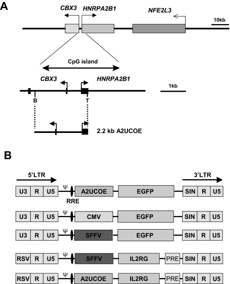
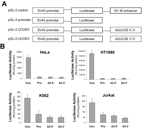
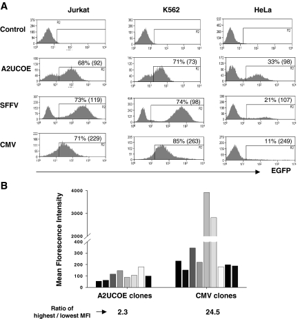
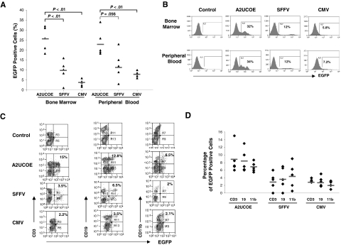
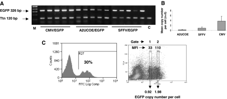
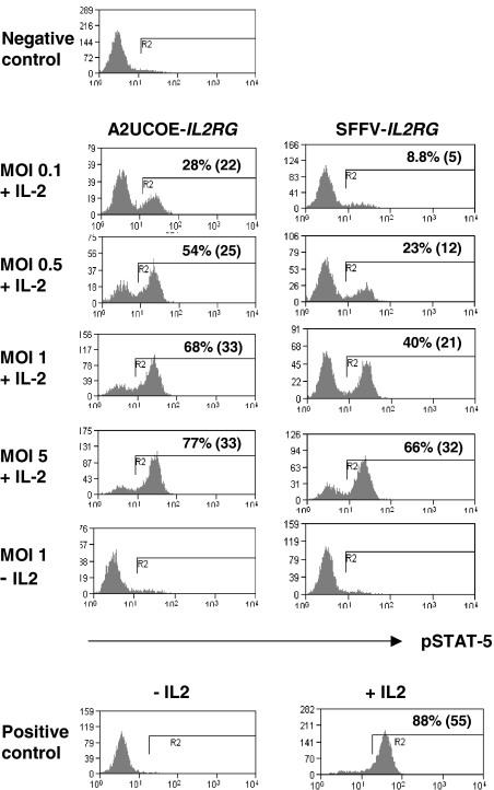
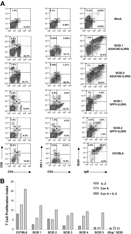
Ubiquitously acting chromatin opening elements (UCOEs) consist of methylation-free CpG islands encompassing dual divergently transcribed promoters of housekeeping genes that have been shown to confer resistance to transcriptional silencing and to produce consistent and stable transgene expression in tissue culture systems. To develop improved strategies for hematopoietic cell gene therapy, we have assessed the potential of the novel human HNRPA2B1-CBX3 UCOE (A2UCOE) within the context of a self-inactivating (SIN) lentiviral vector. Unlike viral promoters, the enhancer-less A2UCOE gave rise to populations of cells that expressed a reporter transgene at a highly reproducible level. The efficiency of expression per vector genome was also markedly increased in vivo compared with vectors incorporating either spleen focus-forming virus (SFFV) or cytomegalovirus (CMV) promoters, suggesting a relative resistance to silencing. Furthermore, an A2UCOE-IL2RG vector fully restored the IL-2 signaling pathway within IL2RG-deficient human cells in vitro and successfully rescued the X-linked severe combined immunodeficiency (SCID-X1) phenotype in a mouse model of this disease. These data indicate that the A2UCOE displays highly reliable transcriptional activity within a lentiviral vector, largely overcoming insertion-site position effects and giving rise to therapeutically relevant levels of gene expression. These properties are achieved in the absence of classic enhancer activity and therefore may confer a high safety profile.


Figures

Figure 1
Figure 1
Illustration of the human HNRPA2B1-CBX3-NFE2L3 locus and derivation of the A2UCOE. (A, top panel) Gene organization of the HNRPA2B1-CBX3-NFE2L3 locus located at chromosome position 7p15.2 (GenBank accession nos. D28877, AC004520, and AC010677) is shown with gene positions indicated (shaded rectangles). (Bottom panel) Expanded view of the 3-kb methylation-free CpG island region encompassing the divergent CBX3 and HNRPA2B1 promoters. The region covered by the minimal 2.2-kb UCOE (A2UCOE; M.A. et al) extending from the TthIII I (T) site within exon I of HNRPA2B1 to a BamHI (B) site within the first intron of CBX3 is shown. Black rectangles denote exons. Horizontal arrows denote the direction of transcription. (B) Illustration of the lentiviral vector constructs. LTR indicates long-terminal repeat; RRE, rev-response element; Ψ, packaging signal; RSV, Rous sarcoma virus U3; and PRE, mutated Woodchuck hepatitis posttranscriptional regulatory element.
Figure 2
Figure 2
The A2UCOE lacks classic enhancer activity. (A) Illustration of the pGL-2 plasmid vector–based constructs. The pGL-2 promoter construct consists of a minimal, enhancer-less SV40 promoter driving a luciferase reporter gene. A 2.2-kb BamHI fragment consisting of a minimal A2UCOE (Figure 1A) was inserted downstream of the luciferase gene in pGL-2 promoter in both forward (pGL-2 UCOE5′) and reverse (pGL-2 UCOE3′) orientations. The pGL-2 control construct has the SV40 enhancer element inserted at the same position as the A2UCOE test fragment and acts as a positive enhancer control. (B) The 4 plasmids illustrated in panel A were used to conduct transient transfection assays in HeLa, HT1080, Jurkat, and K562 cells. Total protein cell lysates were analyzed for luciferase activity 24 hours after transfection. The mean and standard deviation of triplicate experiments for each cell line are shown. Con indicates pGL-2 control; Pro, pGL-2 promoter; A2-5′, pGL-2 A2UCOE5′; and A2-3′, pGL-2 A2UCOE3′. Note that the pGL-2 A2UCOE5′ and pGL-2 A2UCOE3′ test constructs give luciferase activities that are no higher than pGL-2 promoter in all 4 cell lines, indicating the absence of a classic enhancer function within this element.
Figure 3
Figure 3
An A2UCOE regulatory element within a lentiviral vector confers consistent transgene expression. (A) Lentiviral vectors containing an EGFP reporter gene under the control of the A2UCOE, SFFV, or CMV promoters (Figure 1B) were used to transduce Jurkat (T-cell), K562 (myeloid), and HeLa (carcinoma) human cell lines at an MOI of 1. Cells were analyzed by flow cytometry, and plots of percentage EGFP-positive cells (y-axis) verses fluorescence intensity (x-axis) are shown. Data shown are from 14 days of culture. The percentage of total EGFP-positive cells is shown in each plot. The coefficient of variation (CV) of EGFP-positive cells for each type cell is shown in the parentheses. Note that discrete peaks of EGFP-positive cells that are seen with A2UCOE-EGFP in all cells lines in conjunction with a lower CV suggest a more homogeneous transgene expression pattern compared with the SFFV and especially the CMV viral promoters. (B) Lentiviral vectors containing an EGFP reporter gene under control of either the A2UCOE or CMV promoters (Figure 1B) were used to transduce HeLa cells at an MOI of 0.3. Single EGFP-expressing cells were sorted by FACS and clonally expanded in culture. EGFP fluorescence intensity was analyzed by FACS in selected cell clones carrying a single copy of vector transgene. Note that expression from the A2UCOE construct between clones is highly reproducible and 10-fold lower in degree of variation than with the CMV promoter.
Figure 4
Figure 4
The A2UCOE-EGFP vector gives rise to consistent transgene expression in bone marrow and peripheral blood hematopoietic cells in vivo. HSCs were isolated from the bone marrow of C57BL/6J mice and transduced with the A2UCOE-EGFP, SFFV-EGFP, and CMV-EGFP lentiviral vectors (Figure 1B) at an MOI of 20 to 25 and transplanted into lethally irradiated recipient animals. Total peripheral blood and bone marrow cells were obtained at 3 months after transplantation and analyzed for the presence of EGFP-positive cells by FACS. (A) Percentage of total EGFP-positive cells in bone marrow and peripheral blood from A2UCOE, SFFV, and CMV mice (n = 5 in each group). The horizontal bar shown in each column denotes the mean percentage value of EGFP-positive cells in each group. The P values were determined using the Wilcoxon rank sum test. (B) Expression profiles of a representative mouse that received a transplant in each group. The percentage of total EGFP-positive cells is shown in each plot. Note that the A2UCOE-EGFP vector generates a higher percentage of transgene-expressing cells (panel A) as well as discrete peaks of EGFP-positive cell populations (panel B), suggesting a negation of integration-site position effects leading to more consistent transgene expression compared with that achieved with SFFV and CMV viral promoters. (C) Peripheral blood cells from mice transduced with the A2UCOE-, SFFV-, and CMV-EGFP lentiviral vectors were incubated with antibodies against CD3 (T-cell), CD19 (B-cell), and CD11b (myeloid) lineage markers and scored against EGFP expression by FACS analysis. EGFP expression profile in different cell lineages in a representative mouse that received a transplant, corresponding to panel B (bottom row, peripheral blood), in each lentiviral vector group. (D) Total percentage of EGFP-positive cells in T-, B-, and myeloid cell lineages in peripheral blood from A2UCOE, SFFV, and CMV vector–transduced mice (n = 5 in each group). The horizontal bar shown in each column depicts the mean percentage value of EGFP-positive cells in each cell lineage.
Figure 5
Figure 5
A2UCOE-EGFP gives stable, high efficiency expression per vector copy. (A) Genomic DNA derived from bone marrow of mice that received transplants ex vivo of HSCs transduced with the CMV-EGFP, SFFV-EGFP, and A2UCOE-EGFP vectors (Figure 4) was subjected to standard PCR for the presence of transgene (EGFP) and endogenous murine titin (Ttn) sequences and products were resolved by agarose gel electrophoresis. M indicates DNA size markers; C, mock control mouse bone marrow sample. (B) Summary of real-time quantitative PCR analysis of the same samples shown in panel A to determine lentiviral vector copy number. Error bars denote 1 standard deviation about the mean. (C) Determination of vector copy number in subpopulations of EGFP-expressing total bone marrow cells transduced with the A2UCOE-EGFP lentiviral vector. (Left) A representative sample of A2UCOE-EGFP vector–transduced bone marrow cells was sorted by FACS to isolate either low (gate 1) or high (gate 2) EGFP fluorescence intensity cells. DNA was then isolated from the sorted pools of cells and analyzed by QPCR as in panel B. (Right) Profile showing the sorting gates and corresponding mean fluorescence intensity (MFI). Average lentiviral vector copy number per cell is indicated. Note that the A2UCOE gives a higher number of EGFP-positive cells at a lower vector copy number than either the SFFV or CMV promoters (summarized in Table 1), with a clear trend toward copy number–dependent expression.
Figure 6
Figure 6
Rescue of JAK3-mediated STAT-5 tyrosine phosphorylation in human ED-7R cells. The A2UCOE-IL2RG and SFFV-IL2RG lentiviral vectors (Figure 1B) were used to transduce human ED-7R cells at MOIs of 0.1, 0.5, 1, and 5. At 4 days following transduction, cells were stimulated with IL-2, subsequently stained with antiphosphorylated STAT-5 (pSTAT-5) antibody, and assessed for the presence of pSTAT-5 expression by FACS. The percentage of pSTAT-5–positive cells and MFI (in parentheses) is shown. Negative control indicates untransduced ED-7R cells stimulated with IL-2; positive control, ED-7R cells stably transfected with an IL2RG transgene.
Figure 7
Figure 7
Efficient immunologic reconstitution in mice following ex vivo bone marrow HSC gene transfer. (A) HSCs from SCID-X1 mice were transduced with either the A2UCOE-IL2RG or SFFV-IL2RG lentiviral vectors (Figure 1B) and transplanted into lethally irradiated 3KO Il2rg−/−Rag2−/−c5−/− mice (see “Materials and methods, SCID mouse model and ex vivo lentiviral vector–mediated IL2RG gene transfer”). At 3 months following engraftment, spleens were analyzed for reconstitution of T-, B-, and NK-cell lineages by FACS. Cells were stained with anti-CD8, -CD4, -NK1.1, -IgM, and -B220 antibodies. Reconstitution of all cell lineages is observed in A2UCOE-IL2RG vector–transduced mice and SFFV-IL2RG vector–transduced mice (2 SCID-X1 mice transduced with the A2UCOE-IL2RG and 2 transduced with SFFV-IL2RG lentiviral vectors are shown). Mock indicates 3KO; Il2rg−/−Rag2−/−c5−/− SCID-X1 mouse that received only untransduced HSCs. Percentages within quadrants represent the percentage of the total cell population analyzed that is present in that quadrant. (B) T-cell proliferation assay. Splenocytes isolated from mice transduced with A2UCOE-IL2RG were stimulated with Concanavalin A (Con A), IL-2, and Con A plus IL-2. Proliferating cells were assessed by incorporation of 3H-thymidine and expressed as a proliferation index (the ratio of the stimulated cells to unstimulated cells). All 5 mice transduced with A2UCOE-IL2RG showed an increased cell proliferation index compared with an untreated animal.

Similar articles

Cited by

References

    1. Cavazzana-Calvo M, Hacein-Bey S, de Saint BG, et al. Gene therapy of human severe combined immunodeficiency (SCID)-X1 disease. Science. 2000;288:669–672. - PubMed
    1. Aiuti A, Slavin S, Aker M, et al. Correction of ADA-SCID by stem cell gene therapy combined with nonmyeloablative conditioning. Science. 2002;296:2410–2413. - PubMed
    1. Gaspar HB, Parsley KL, Howe S, et al. Gene therapy of X-linked severe combined immunodeficiency by use of a pseudotyped gammaretroviral vector. Lancet. 2004;364:2181–2187. - PubMed
    1. Gaspar HB, Bjorkegren E, Parsley K, et al. Successful reconstitution of immunity in ADA-SCID by stem cell gene therapy following cessation of PEG-ADA and use of mild preconditioning. Mol Ther. 2006;14:505–513. - PubMed
    1. Schmidt M, Hacein-Bey-Abina S, Wissler M, et al. Clonal evidence for the transduction of CD34(+) cells with lymphomyeloid differentiation potential and self-renewal capacity in the SCID-X1 gene therapy trial. Blood. 2005;105:2699–2706. - PubMed

Publication types

MeSH terms