Case Reports
. 2007 Jul;121(6):697-709.
doi: 10.1007/s00439-007-0359-6. Epub 2007 Apr 25.

Complex chromosome 17p rearrangements associated with low-copy repeats in two patients with congenital anomalies

Affiliations
Case Reports

Complex chromosome 17p rearrangements associated with low-copy repeats in two patients with congenital anomalies

L E L M Vissers et al. Hum Genet. 2007 Jul.

Abstract

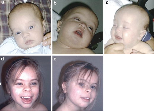
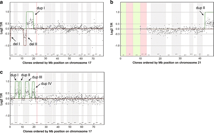
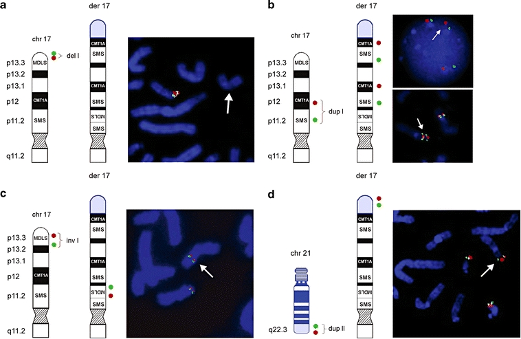
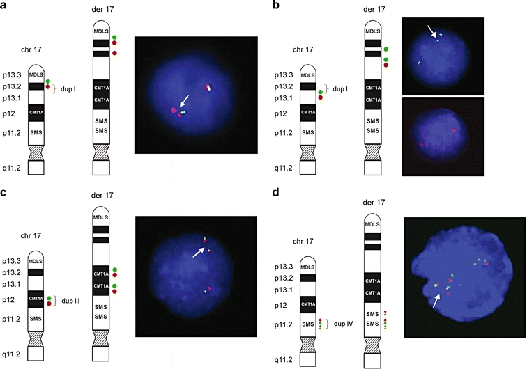
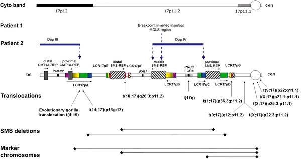
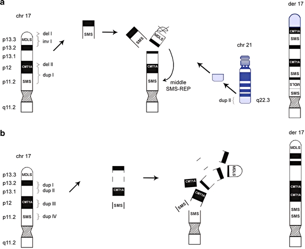
Recent molecular cytogenetic data have shown that the constitution of complex chromosome rearrangements (CCRs) may be more complicated than previously thought. The complicated nature of these rearrangements challenges the accurate delineation of the chromosomal breakpoints and mechanisms involved. Here, we report a molecular cytogenetic analysis of two patients with congenital anomalies and unbalanced de novo CCRs involving chromosome 17p using high-resolution array-based comparative genomic hybridization (array CGH) and fluorescent in situ hybridization (FISH). In the first patient, a 4-month-old boy with developmental delay, hypotonia, growth retardation, coronal synostosis, mild hypertelorism, and bilateral club feet, we found a duplication of the Charcot-Marie-Tooth disease type 1A and Smith-Magenis syndrome (SMS) chromosome regions, inverted insertion of the Miller-Dieker lissencephaly syndrome region into the SMS region, and two microdeletions including a terminal deletion of 17p. The latter, together with a duplication of 21q22.3-qter detected by array CGH, are likely the unbalanced product of a translocation t(17;21)(p13.3;q22.3). In the second patient, an 8-year-old girl with mental retardation, short stature, microcephaly and mild dysmorphic features, we identified four submicroscopic interspersed 17p duplications. All 17 breakpoints were examined in detail by FISH analysis. We found that four of the breakpoints mapped within known low-copy repeats (LCRs), including LCR17pA, middle SMS-REP/LCR17pB block, and LCR17pC. Our findings suggest that the LCR burden in proximal 17p may have stimulated the formation of these CCRs and, thus, that genome architectural features such as LCRs may have been instrumental in the generation of these CCRs.


Figures

Fig. 1
Fig. 1
Patient 1 (ac) at the age of 4 months. Patient 2 (de) at the age of 6 years 10 months
Fig. 2
Fig. 2
Chromosome 17 (patient 1 and 2) and chromosome 21 (patient 1) array CGH profiles. On the x-axis, clones are ordered by Mb position on chromosome 17 and 21, respectively, and on the y-axis log2T/R ratios are shown. Hidden Markov Model was used to identify the duplications (green lines) and deletions (red lines) in patient 1 and 2. a In patient 1, chromosome 17 shows two interspersed deletions of ∼600 kb (17p13.3–Del I) and ∼2.2 Mb (17p12–Del II), respectively, and a duplication of ∼6.1 Mb (Dup I). b Additionally, a 4.4 Mb duplication of 21q22.3 was observed (Dup II). c Patient 2 showed four interspersed duplications on 17p11–p13 (Dup I–Dup IV), in total comprising ∼8.8 Mb of genomic sequence. All alterations were shown to be de novo
Fig. 3
Fig. 3
Ideograms and FISH results of patient 1. Schematic representation of a normal 17p and der(17) (black and white) with translocated chromosome 21 material (blue). The location of the FISH probes are shown on the left side of each figure panel; der(17) is indicated on FISH pictures by a white arrow. a Terminal deletion of 17pter was validated using BAC clones RP11-1260E13 (red) and CTD-2326F1 (green) (del I). b FISH with PMP22-specific PAC RP1-150M12 (red) RA11-specific and BAC RP11-525O11 (green) revealed direct duplication of the CMT1A and SMS regions in 17p12p11.2 (dup I). c FISH with PAC RP1-95H6 (red; adapted from Chong et al. 1997) and BAC GS-202L17 (green; adapted from Knight et al. 2000) showed inverted insertion of the MDLS region into the SMS region. d Array CGH also identified a duplication of 21q22.3 (dup II). Additional FISH analysis using BAC clones RP11-40L10 (green) and RP11-16B19 (red) revealed that the duplicated material of 21q22.3 was translocated onto der(17). Summary of FISH results is provided in Table 1
Fig. 4
Fig. 4
Ideograms and FISH results of patient 2. Schematic representation of the normal 17p and der(17). The locations of the FISH probes are shown on the left side of each figure panel. a The distal breakpoint of duplication I showed a relatively simple fluorescence signal pattern with probes RP11-810M2 (green; normal) and RP11-597I9 (red; duplicated). b The proximal breakpoint of duplication I showed a duplicated signal for RP11-222J21 (green) and a normal signal for RP11-98D15 (red). c Direct orientation of duplication III was shown using BAC clones RP11-601N13 (green) and RP11-726O12 (red). d For the distal breakpoint of duplication IV, BAC clones RP11-448D22 (green) and CTD-2145A24 (red) showed duplicated signals on der(17), indicating that both middle SMS-REP and LCR17pB are duplicated as a block. Four red signals on der(17) representing two normal and two duplicated copies of LCR17pA/B (dup III) and LCR17pB (dup IV) and four green signals depicting three normal copies of SMS-REPs and the duplicated middle SMS-REP. Summary of FISH results is provided in Table 2
Fig. 5
Fig. 5
Schematic diagram of breakpoints for DNA rearrangements in 17p. Top: Cytogenetic chromosome bands 17p12, and sub-bands 17p11.2 and 17p11.1 are shown. Breakpoints of the chromosome rearrangements in patients 1 and 2 associated with architectural features in proximal 17p are shown by vertical dotted blue arrows. In patient 1, one breakpoint is located within the middle SMS-REP/LCR17pB block, whereas in patient 2 breakpoints are located within LCR17pA (proximal Dup III), middle SMS-REP (distal Dup IV) and LCR17pC (proximal Dup IV). Duplication III and IV in patient 2 are indicated by horizontal blue lines. Bottom: Previously identified rearrangements associated with LCRs in 17p. Breakpoints of translocations and isochromosome 17q are indicated by vertical black arrows whereas common ∼4 Mb and uncommon ∼5 Mb SMS deletions and marker chromosomes are indicated by black horizontal lines. The LCR17p structures are depicted in colors to better represent their positional orientation with respect to each other; the shaded rectangles and horizontal black arrows represent the orientation of the LCRs
Fig. 6
Fig. 6
Schematic representation of the proposed mechanism for CCR formation in patient 1 and patient 2. a The complex rearrangement observed in patient 1 includes nine breakpoints, with one insertion, two microdeletions and two microduplications. We propose that the breaks in chromosome 17, p13.3, p12 and p11.2 as well as the break in chromosome 21q22.3 arose simultaneously. This resulted in the insertion of the MDLS region into the middle SMS-REP/LCR17pB block, loss of telomeric 17p and part of 17p12 and duplication of the CMT1A region. Additionally, the duplicated 21q22.3 fragment was translocated to the 17p subtelomeric region. b The complex rearrangement in patient 2 included eight breaks. We suggest that breaks in chromosome 17, p13.2, p13.1, p12 and p11.2 occurred at the same time, resulting in four interspersed directly orientated microduplications. Arrows do not represent a chronological order of events

Similar articles

Cited by

References

    1. {'text': '', 'ref_index': 1, 'ids': [{'type': 'DOI', 'value': '10.1007/s00439-003-1079-1', 'is_inner': False, 'url': 'https://doi.org/10.1007/s00439-003-1079-1'}, {'type': 'PubMed', 'value': '14767757', 'is_inner': True, 'url': 'https://pubmed.ncbi.nlm.nih.gov/14767757/'}]}
    2. Astbury C, Christ LA, Aughton DJ, Cassidy SB, Fujimoto A, Pletcher BA, Schafer IA, Schwartz S (2004) Delineation of complex chromosomal rearrangements: evidence for increased complexity. Hum Genet 114:448–457 - PubMed
    1. {'text': '', 'ref_index': 1, 'ids': [{'type': 'DOI', 'value': '10.1038/ng0306-269', 'is_inner': False, 'url': 'https://doi.org/10.1038/ng0306-269'}, {'type': 'PubMed', 'value': '16940994', 'is_inner': True, 'url': 'https://pubmed.ncbi.nlm.nih.gov/16940994/'}]}
    2. Axton M (2006) Structural variants deconstruct the genome. Nat Genet 38:959 - PubMed
    1. {'text': '', 'ref_index': 1, 'ids': [{'type': 'DOI', 'value': '10.1101/gr.GR-1871R', 'is_inner': False, 'url': 'https://doi.org/10.1101/gr.gr-1871r'}, {'type': 'PMC', 'value': 'PMC311093', 'is_inner': False, 'url': 'https://pmc.ncbi.nlm.nih.gov/articles/PMC311093/'}, {'type': 'PubMed', 'value': '11381028', 'is_inner': True, 'url': 'https://pubmed.ncbi.nlm.nih.gov/11381028/'}]}
    2. Bailey JA, Yavor AM, Massa HF, Trask BJ, Eichler EE (2001) Segmental duplications: organization and impact within the current human genome project assembly. Genome Res 11:1005–1017 - PMC - PubMed
    1. {'text': '', 'ref_index': 1, 'ids': [{'type': 'DOI', 'value': '10.1126/science.1072047', 'is_inner': False, 'url': 'https://doi.org/10.1126/science.1072047'}, {'type': 'PubMed', 'value': '12169732', 'is_inner': True, 'url': 'https://pubmed.ncbi.nlm.nih.gov/12169732/'}]}
    2. Bailey JA, Gu Z, Clark RA, Reinert K, Samonte RV, Schwartz S, Adams MD, Myers EW, Li PW, Eichler EE (2002) Recent segmental duplications in the human genome. Science 297:1003–1007 - PubMed
    1. Bartels I, Starke H, Argyriou L, Sauter SM, Zoll B, Liehr T (2006) An exceptional complex chromosomal rearrangement (CCR) with eight breakpoints involving four chromosomes (1;3;9;14) in an azoospermic male with normal phenotype. Eur J Med Genet doi:10.1016/j.ejmg.2006.10.007 - PubMed

Publication types

LinkOut - more resources