Comparative Study
. 2007 May;57(5):881-90.
doi: 10.1002/mrm.21176.

Clinical multishot DW-EPI through parallel imaging with considerations of susceptibility, motion, and noise

Affiliations
Comparative Study

Clinical multishot DW-EPI through parallel imaging with considerations of susceptibility, motion, and noise

Stefan Skare et al. Magn Reson Med. 2007 May.

Abstract

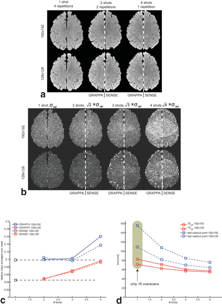
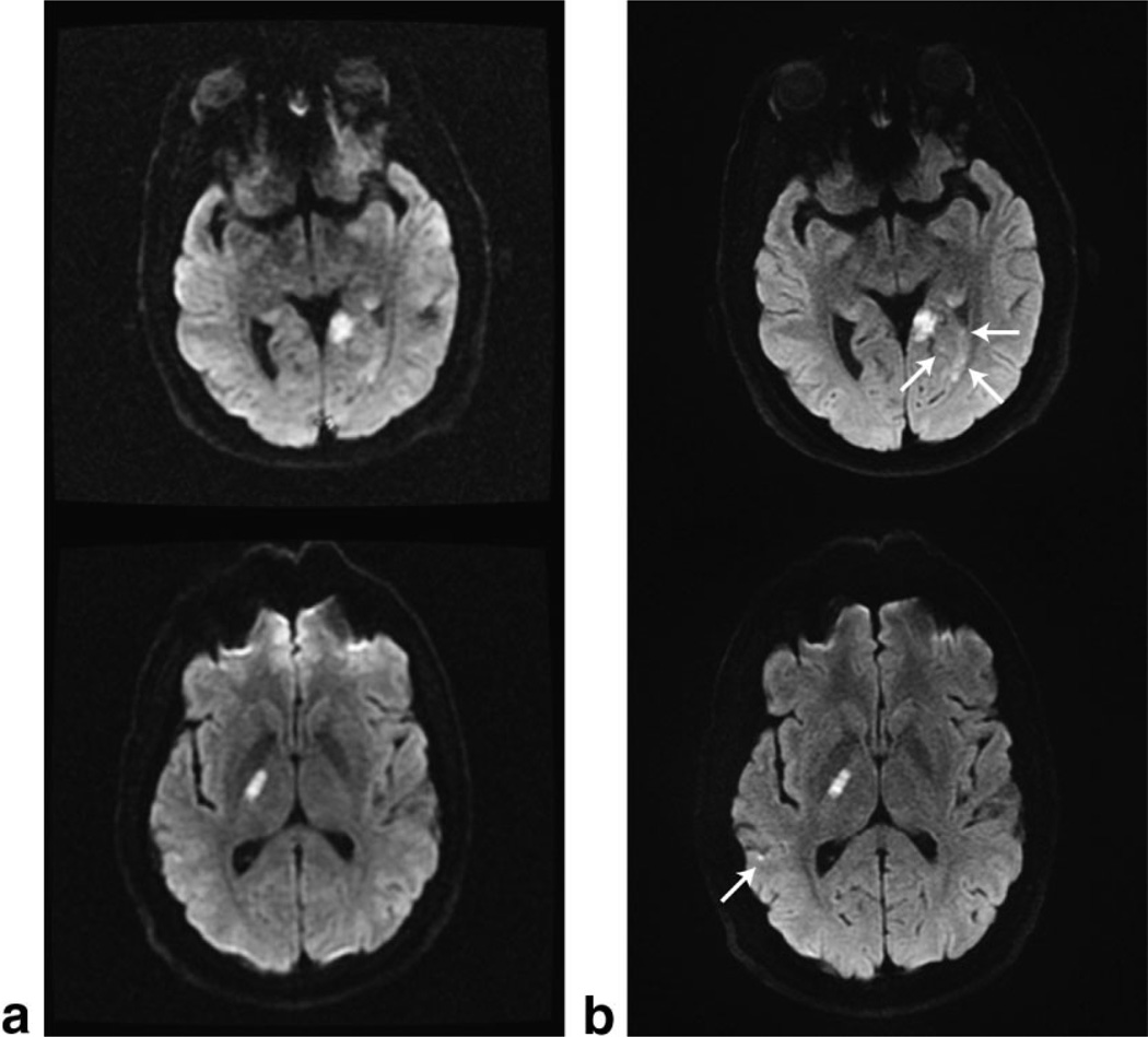
Geometric distortions and poor image resolution are well known shortcomings of single-shot echo-planar imaging (ss-EPI). Yet, due to the motion immunity of ss-EPI, it remains the most common sequence for diffusion-weighted imaging (DWI). Moreover, both navigated DW interleaved EPI (iEPI) and parallel imaging (PI) methods, such as sensitivity encoding (SENSE) and generalized autocalibrating parallel acquisitions (GRAPPA), can improve the image quality in EPI. In this work, DW-EPI accelerated by PI is proposed as a self-calibrated and unnavigated form of interleaved acquisition. The PI calibration is performed on the b = 0 s/mm2 data and applied to each shot in the rest of the DW data set, followed by magnitude averaging. Central in this study is the comparison of GRAPPA and SENSE in the presence of off-resonances and motion. The results show that GRAPPA is more robust than SENSE against both off-resonance and motion-related artifacts. The SNR efficiency was also investigated, and it is shown that the SNR/scan time ratio is equally high for one- to three-shot high-resolution diffusion scans due to the shortened EPI readout train length. The image quality improvements without SNR efficiency loss, together with motion tolerance, make the GRAPPA-driven DW-EPI sequence clinically attractive.


Figures

FIG. 1
FIG. 1
Conventional self-calibrated GRAPPA in combination with EPI. (a) ss-EPI and (c) conventional SE show the phantom with and without geometric distortions. Using a variable k-space sampling scheme for EPI with an ORF of 4 results in a GRAPPA-reconstructed image (b). Here the low-resolution portion of the image remains shifted, whereas the higher spatial frequencies (edges) are four times less distorted than the low-spatial-frequency components, yielding this double-image appearance. The white dashed lines are guides for the true phantom edges defined in c and the distorted edges in a. This is evidence that conventional self-calibration may not be used with EPI.
FIG. 2
FIG. 2
3T volunteer data showing that external calibration data (ac) with different amounts of distortion affect the image quality for GRAPPA (df) and SENSE (gi) reconstructions of three-shot DWI data. (a) Conventional SE, (b) three-shot b0 EPI, (c) one-shot b0 EPI, (d–f) GRAPPA-reconstructed, and (g–i) SENSE-reconstructed three-shot isotropic DW images using calibration data are shown on the same row. Overall, both GRAPPA and SENSE show little dependence on the choice of calibration data, except for the missing data for SENSE in i and slight noise enhancement in f (see insets for f and i). This gives flexibility in the choice of calibration data, and shows that the proposed use of a multishot b0 image for calibration purposes is not suboptimal compared to, e.g., a time-consuming SE calibration scan (a).
FIG. 3
FIG. 3
a: Three-shot b0 EPI data acquired with the head rotated 40°. The contours of the following three-shot isotropic DWI are outlined in red. The data in a are reconstructed with (b) GRAPPA and (c) SENSE. GRAPPA is shown to be immune to motion that may occur between the acquisition of the data used for calibration and the subsequent DWI. [Color figure can be viewed in the online issue, which is available at www.interscience.wiley.com.]
FIG. 4
FIG. 4
Sustained motion during the entire diffusion scan (i.e., also during the acquisition of the first b0 image used for PI calibration). a: b0 image reconstructed without PI and used for the estimation of coil sensitivity maps and GRAPPA weights. b: Same b0 image, where each of the three shots has been reconstructed using the GRAPPA weights obtained from the image itself, followed by motion correction between the “shot-images” and magnitude averaging. Isotropic DWI using the same GRAPPA weights, (c) without and (d) with motion correction between the shots, is shown. The corresponding SENSE reconstructions are shown in e–g. Unlike GRAPPA, the SENSE reconstruction is impaired by the intershot ghosts in a, which on top of residual aliasing in the brain also deceives the intensity-based motion correction.
FIG. 5
FIG. 5
SNR efficiency of volunteer brain data. a: Split view with both GRAPPA and SENSE reconstructions in each subpanel for an easy noise comparison between the two for different image resolutions and shots. All data have been averaged over different numbers of repetitions to normalize by scan time. One and two shots are comparable, while the structured noise is disturbing for the four-shot case. b: Relative SD maps, normalized to the same scan time (σrelNinter, Eq. [1]). For 192 × 192, there is no apparent noise difference between one to three shots. In c the mean of σrelNinter over all brain pixels in b is given. The black dots and dashed lines indicate the noise level of the ss-EPI, which is constant for one- to three-shot GRAPPA with a 192 × 192 matrix. In d the TEs and time for the end of the EPI train are given. Overall, SENSE and GRAPPA demonstrate very similar noise characteristics, except for 192 × 192 at R = 4, where SENSE has a small advantage.
FIG. 6
FIG. 6
Clinical examples of the standard diffusion sequence using (a) 128 × 128 ss-EPI data acquired in less than 1 min, and (b) our three-shot 192 × 192 DW-EPI data reconstructed with GRAPPA using a tetrahedral diffusion scheme repeated three times. The top and bottom rows were acquired with the eight-channel neurovascular array coil and the eight-channel brain coil, respectively.

References

    1. Moseley ME, Kucharczyk J, Mintorovitch J, Cohen Y, Kurhanewicz J, Derugin N, Asgari H, Norman D. Diffusion-weighted MR imaging of acute stroke: correlation with T2-weighted and magnetic susceptibility-enhanced MR imaging in cats. AJNR Am J Neuroradiol. 1990;11:423–429. - PMC - PubMed
    1. Ebisu T, Tanaka C, Umeda M, Kitamura M, Naruse S, Higuchi T, Ueda S, Sato H. Discrimination of brain abscess from necrotic or cystic tumors by diffusion-weighted echo planar imaging. Magn Reson Imaging. 1996;14:1113–1116. - PubMed
    1. Kim YJ, Chang KH, Song IC, Kim HD, Seong SO, Kim YH, Han MH. Brain abscess and necrotic or cystic brain tumor: discrimination with signal intensity on diffusion-weighted MR imaging. AJR Am J Roentgenol. 1998;171:1487–1490. - PubMed
    1. Lai PH, Ho JT, Chen WL, Hsu SS, Wang JS, Pan HB, Yang CF. Brain abscess and necrotic brain tumor: discrimination with proton MR spectroscopy and diffusion-weighted imaging. AJNR Am J Neuroradiol. 2002;23:1369–1377. - PMC - PubMed
    1. Farzaneh F, Riederer SJ, Pelc NJ. Analysis of T2 limitations and off-resonance effects on spatial resolution and artifacts in echo-planar imaging. Magn Reson Med. 1990;14:123–139. - PubMed

Publication types

MeSH terms