Review
. 2007 Mar 7;13(9):1335-46.
doi: 10.3748/wjg.v13.i9.1335.

Function of longitudinal vs circular muscle fibers in esophageal peristalsis, deduced with mathematical modeling

Affiliations
Review

Function of longitudinal vs circular muscle fibers in esophageal peristalsis, deduced with mathematical modeling

James G Brasseur et al. World J Gastroenterol. .

Abstract

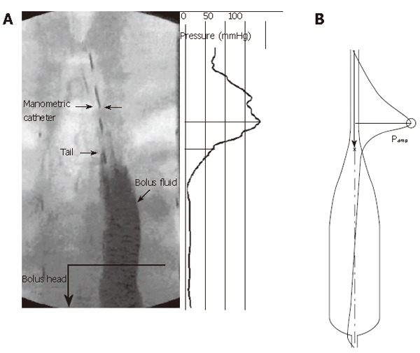
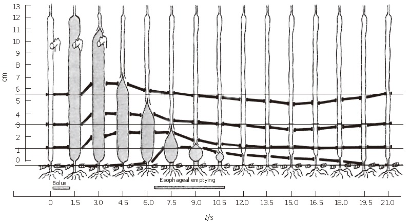
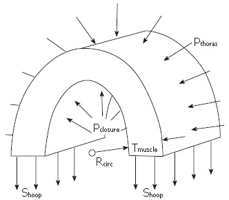
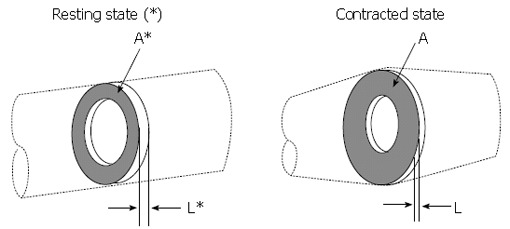
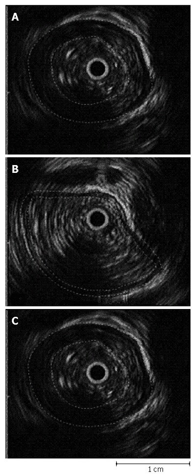
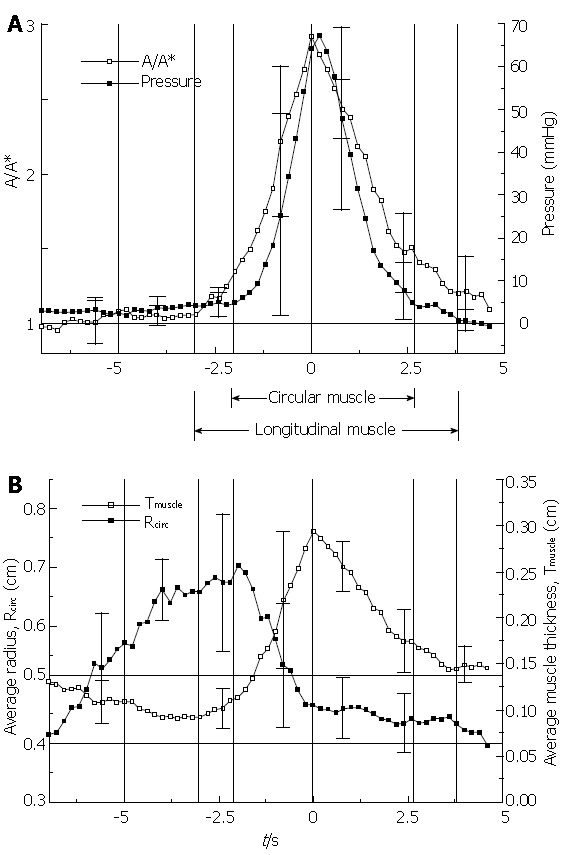
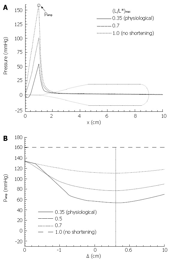
We summarize from previous works the functions of circular vs. longitudinal muscle in esophageal peristaltic bolus transport using a mix of experimental data, the conservation laws of mechanics and mathematical modeling. Whereas circular muscle tone generates radial closure pressure to create a local peristaltic closure wave, longitudinal muscle tone has two functions, one physiological with mechanical implications, and one purely mechanical. Each of these functions independently reduces the tension of individual circular muscle fibers to maintain closure as a consequence of shortening of longitudinal muscle locally coordinated with increasing circular muscle tone. The physiological function is deduced by combining basic laws of mechanics with concurrent measurements of intraluminal pressure from manometry, and changes in cross sectional muscle area from endoluminal ultrasound from which local longitudinal shortening (LLS) can be accurately obtained. The purely mechanical function of LLS was discovered from mathematical modeling of peristaltic esophageal transport with the axial wall motion generated by LLS. Physiologically, LLS concentrates circular muscle fibers where closure pressure is highest. However, the mechanical function of LLS is to reduce the level of pressure required to maintain closure. The combined physiological and mechanical consequences of LLS are to reduce circular muscle fiber tension and power by as much as 1/10 what would be required for peristalsis without the longitudinal muscle layer, a tremendous benefit that may explain the existence of longitudinal muscle fiber in the gut. We also review what is understood of the role of longitudinal muscle in esophageal emptying, reflux and pathology.


Figures

Figure 1
Figure 1
Schematic of esophageal cross section, showing the primary esophageal layers.
Figure 2
Figure 2
Frozen time image of peristaltic transport of a liquid bolus through the esophagus, with corresponding spatial variation in intraluminal pressure. A: Fluoroscopic image and pressure distribution (relative to atmospheric) concurrent with interpolated high-resolution manometry data; B: Bolus shape and overlayed pressure distribution from a mathematical model computer simulation.
Figure 3
Figure 3
The classic experiment by Dodds et al[10] in which the motion of four material points in the muscle wall of the feline esophagus were recorded over time concurrently with bolus transport using fluoroscopy.
Figure 4
Figure 4
Schematic of the force balance across an axial segment of the muscularis propria of the esophageal wall, showing the relationship between closure pressure (Pclosure) and average hoop stress (Shoop) across the circular and longitudinal muscle layers.
Math 1
Math 1
Math(A1).
Figure 5
Figure 5
Application of the principle of mass conservation to quantify local longitudinal shortening from measurement of the change in cross sectional area from endoluminal ultrasound images, leading to L/L* = 1/(A/A*).
Math 2
Math 2
Math(A2).
Figure 6
Figure 6
Example of the use of EUS with image analysis to determine the cross sectional area of the muscularis propria (A) in the resting state (*), (B) with distention at the bolus head, and (C) at peak contractile pressure.
Figure 7
Figure 7
Schematic of essential elements in the mathematical model. A: Bolus shape with geometrical parameters. H is the bolus head radius, λ is bolus length, ε is the thickness of the lubrication layer in the contracted zone, and c is the peristaltic wave speed; B: Specification of mucosal surface velocity in the model. Ub and Vb are the axial and radial surface velocity components, respectively, of a material element of the mucosal surface.
Figure 8
Figure 8
The primary result from Nicosia et al[1]. In (A) the inverse of local longitudinal shortening (A/A* = 1/(L/L*), see equation 2) is plotted together with circular muscle closure pressure (see equation 1) and in (B) the effective thickness of the muscularis propria is plotted together with effective lumen radius. All variables are plotted during the passage of a peristaltic wave with the transport of a 10 mL liquid bolus in the mid esophagus. Averages over four normal subjects were done referenced to the peak in LLS.
Figure 9
Figure 9
From Dai et al[2], local longitudinal shortening at peak intraluminal pressure during swallowing of 5 mL water bolus at different locations above the upper margin of lower esophageal sphincter high-pressure zone. Averages of 20 normal subjects.
Figure 10
Figure 10
Schematic of the parameterization of local longitudinal shortening in the model. The shape of the shortening parameter L/L* is fixed while maximum LLS and offset between the longitudinal and circular muscle contraction waves are varied.
Figure 11
Figure 11
Primary result from Pal & Brasseur[18]. A: Mathematical model calculation of pressure during esophageal peristaltic transport with different levels of local longitudinal shortening, from the measured physiological level, to no shortening; B: Calculation of peak closure pressure as a function of separation △ between the circular muscle and longitudinal muscle contraction waves (Figure 10).

Similar articles

Cited by

References

    1. Nicosia MA, Brasseur JG, Liu JB, Miller LS. Local longitudinal muscle shortening of the human esophagus from high-frequency ultrasonography. Am J Physiol Gastrointest Liver Physiol. 2001;281:G1022–G1033. - PubMed
    1. Dai Q, Korimilli A, Thangada VK, Chung CY, Parkman H, Brasseur J, Miller LS. Muscle shortening along the normal esophagus during swallowing. Dig Dis Sci. 2006;51:105–109. - PubMed
    1. Ulerich R, Dai Q, Miller LS, Brasseur JG. Detailed 3-D Anatomy of the Human Gastro-Esophageal Segment. Gastroenterology. 2003;124:A259–A259.
    1. Liu JB, Miller LS, Goldberg BB, Feld RI, Alexander AA, Needleman L, Castell DO, Klenn PJ, Millward CL. Transnasal US of the esophagus: preliminary morphologic and function studies. Radiology. 1992;184:721–727. - PubMed
    1. Miller LS, Liu JB, Colizzo FP, Ter H, Marzano J, Barbarevech C, Helwig K, Leung L, Goldberg BB, Hedwig K corrected to Helwig K. Correlation of high-frequency esophageal ultrasonography and manometry in the study of esophageal motility. Gastroenterology. 1995;109:832–837. - PubMed

MeSH terms