Review
. 2007 Mar 7;13(9):1408-21.
doi: 10.3748/wjg.v13.i9.1408.

Advanced imaging and visualization in gastrointestinal disorders

Affiliations
Review

Advanced imaging and visualization in gastrointestinal disorders

Odd Helge Gilja et al. World J Gastroenterol. .

Abstract

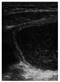
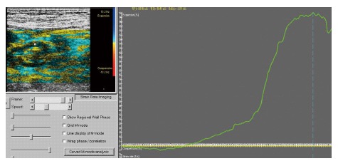
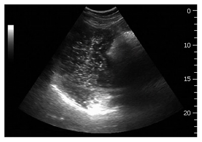
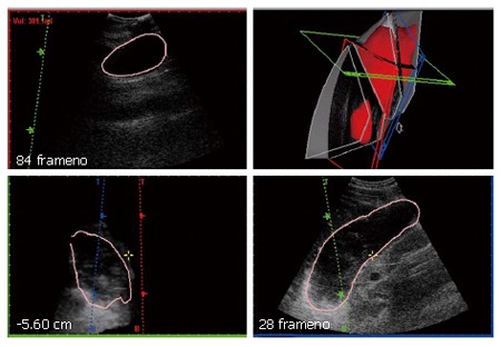
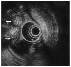
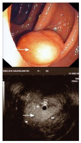
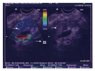
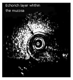
Advanced medical imaging and visualization has a strong impact on research and clinical decision making in gastroenterology. The aim of this paper is to show how imaging and visualization can disclose structural and functional abnormalities of the gastrointestinal (GI) tract. Imaging methods such as ultrasonography, magnetic resonance imaging (MRI), endoscopy, endosonography, and elastography will be outlined and visualization with Virtual Reality and haptic methods. Ultrasonography is a versatile method that can be used to evaluate antral contractility, gastric emptying, transpyloric flow, gastric configuration, intragastric distribution of meals, gastric accommodation and strain measurement of the gastric wall. Advanced methods for endoscopic ultrasound, three-dimensional (3D) ultrasound, and tissue Doppler (Strain Rate Imaging) provide detailed information of the GI tract. Food hypersensitivity reactions including gastrointestinal reactions due to food allergy can be visualized by ultrasonography and MRI. Development of multi-parametric and multi-modal imaging may increase diagnostic benefits and facilitate fusion of diagnostic and therapeutic imaging in the future.


Figures

Figure 1
Figure 1
Ultrasonogram of the fluid-filled gastric antrum showing the different wall layers and a propagating contraction wave that occludes the lumen.
Figure 2
Figure 2
These graphics outline the relative radial strain of the circular muscle layer of the antral wall. The exact sampling point in the gastric wall is denoted with a red marker in the color Doppler ultrasonogram of the left panel. In the right panel, the positive strain curve with a maximum of 150% radial elongation of the muscle layer is demonstrated.
Figure 3
Figure 3
An oblique frontal section applied for ultrasound scanning of the proximal stomach. The top margin of the fundus is shown as a white line in the bottom of the image. A proximal gastric diameter is outlined in the frontal section by tracing normally to the longitudinal axis of the proximal stomach, thus indicating the size of the proximal stomach.
Figure 4
Figure 4
A 3D reconstruction of the total stomach volume (red) in the upper left panel. The acquisition is based on magnetic scanhead tracking. Three orthogonal orientation planes are shown in red, green and blue. The upper left panel is a volume reconstruction window where manual outlining of the structures are made.
Figure 5
Figure 5
Small bowel loops (arrows) imaged from the stomach with an echoendoscope. Liq-uid within the gastric lumen gives an excellent acoustic window.
Figure 6
Figure 6
Endoscopic image of a tumour in the colon (arrow, top). An ultrasound miniprobe is used to “palpate” the relatively soft echo-rich tumour (arrow) which is a lipoma.
Figure 7
Figure 7
ES-elastography (colour coded, left) in chronic pancreatitis displaying hard (blue) and softer (green) areas (left, large arrow). Corresponding B-image (right, arrow). Transducer pressure is indicated by a scale (small arrow).
Figure 8
Figure 8
Endosonographic changes i.e. any resolvable wall thickening of the duodenum, contraction or new echogenic layer) were observed in many patients with food hypersensitivity. Increased mucosal thickness associated with a new echogenic layer was seen in two patients[82].
Figure 9
Figure 9
The sonographic features; wall thickness and diameter of the duodenal bulb and jejunum, peristalsis activity, and luminal fluid were recorded before and during allergen challenge by external ultrasound. The ultrasonographic view of the duodenal bulb shows wall thickening and increased diameter in response to provocation[83].
Figure 10
Figure 10
Example of a colonoscopy layout in a 3D medical workstation 3Dnet Examiner (Copyright Biotronics3D Ltd, Used by permission).
Figure 11
Figure 11
Comparison between real colon dissection and the resulting visualization of colon unfolding technique (Copyright Anna Vilanova, Used by permission).
Figure 12
Figure 12
Screenshot of current two-dimensional ultrasound elastography visualization (A) and the Novint Falcon 3D haptic controller (B).

Similar articles

Cited by

References

    1. Gilja OH, Hausken T, Ødegaard S, Wendelbo Ø, Thierley M. Mobile ultrasonography in a medical department. Tidsskr Nor Laegeforen. 2003;123:2713–2714. - PubMed
    1. Bateman DN, Leeman S, Metreweli C, Willson K. A non-invasive technique for gastric motility measurement. Br J Radiol. 1977;50:526–527. - PubMed
    1. Holt S, McDicken WN, Anderson T, Stewart IC, Heading RC. Dynamic imaging of the stomach by real-time ultrasound--a method for the study of gastric motility. Gut. 1980;21:597–601. - PMC - PubMed
    1. Hausken T, Berstad A. Wide gastric antrum in patients with non-ulcer dyspepsia. Effect of cisapride. Scand J Gastroenterol. 1992;27:427–432. - PubMed
    1. Hausken T, Odegaard S, Berstad A. Antroduodenal motility studied by real-time ultrasonography. Effect of enprostil. Gastroenterology. 1991;100:59–63. - PubMed

MeSH terms