. 2007 Jul;98(7):1027-34.
doi: 10.1111/j.1349-7006.2007.00494.x. Epub 2007 Apr 24.

Occludin-mediated premature senescence is a fail-safe mechanism against tumorigenesis in breast carcinoma cells

Affiliations

Occludin-mediated premature senescence is a fail-safe mechanism against tumorigenesis in breast carcinoma cells

Makoto Osanai et al. Cancer Sci. 2007 Jul.

Abstract

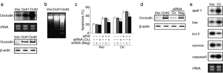
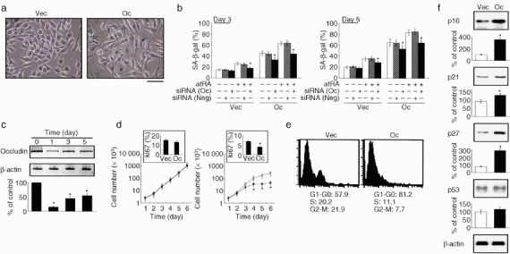
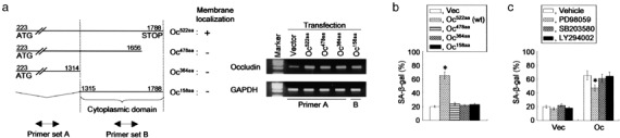
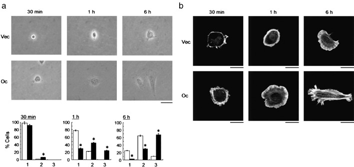
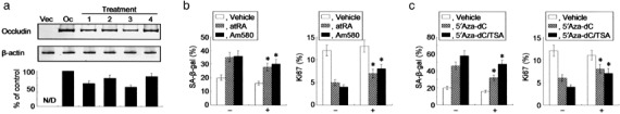
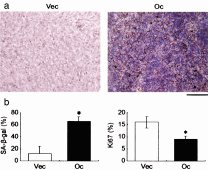
We have previously demonstrated that epigenetic silencing of occludin, a tight junction-associated membrane protein, results in the acquisition of apoptotic resistance to various apoptogenic stimuli, causally contributing to the enhanced tumorigenicity of cancer cells. However, it remains to be examined whether occludin expression in transformed cells has an alternative impact that is important for cancer progression. Here we show that forced expression of occludin induces anoikis and promotes oxidative stress-induced premature senescence in breast carcinoma cells, which is accompanied by upregulation of negative cell cycle regulators such as p16(INK4A), p21(Waf1/Cip1) and p27(Kip1) but not p53. The senescent phenotype is reversed by specific inhibition of mitogen-activated protein kinase. Endogenous reexpression of occludin mediated by a synergistic effect with a demethylator and histone deacetylase inhibitor or retinoids that stimulate retinoic acid receptor alpha is also sufficient for provoking the senescent phenotype. In addition, tumors that developed from occludin-expressing cells in mice showed a feature of cellular senescence that has not been described as a consequence of occludin signaling. These findings suggest that the loss of occludin expression is at least partially involved in the senescence-escape program during mammary tumorigenesis.


Figures

Figure 1
Figure 1
Forced expression of occludin results in enhanced sensitivity to anoikis in AC2M2 cells. (a) Reverse transcription–polymerase chain reaction (RT‐PCR) and Southern blot analysis (upper panel) and western blot analysis (lower panel) to show the expression of occludin in AC2M2‐Vec (Vec), and constitutively overexpressed wild‐type occludin in AC2M2‐Oc#1 (Oc#1) and AC2M2‐Oc#2 (Oc#2) cells. (b,c) Anoikis was induced after adding cells to agarose‐coated dishes to avoid cell attachment. DNA laddering analysis to confirm apoptotic cell death (b). Quantification of anoikis in the presence or absence of 1 µM all‐trans retinoic acid as an apoptotic sensitizer in combination with or without occludin‐specific (Oc) or negative control (Neg) small interfering RNA (siRNA) as determined by terminal deoxynucleotidyl transferase‐meditated dUTP nick‐end labeling assay (c). siRNA (100 nM) was transfected 48 h before anoikis induction. *P < 0.05 versus cells without siRNA transfection. (d) Western blot analysis confirmed the silencing effect of occludin‐specific siRNA (Oc) in AC2M2‐Oc#2 cells (Oc#2), but negative control siRNA (Neg) had no effect on the cells. (e) Occludin expression affects gene expression involving apoptotic machinery. RT‐PCR and Southern blot analysis of indicated apoptosis‐associated genes in AC2M2‐Vec (Vec) and AC2M2‐Oc#2 (Oc) cells.
Figure 2
Figure 2
Occludin induces premature senescence in AC2M2 cells under oxidative stress. (a,b) AC2M2‐Vec (Vec) and AC2M2‐Oc (Oc) cells were exposed to 25 µM H2O2 for up to 5 days in the presence or absence of 1 µM all‐trans retinoic acid in combination with or without 100 nM occludin‐specific (Oc) or negative control (Neg) small interfering RNA (siRNA), and were stained for SA‐β‐gal. Note the blue cytoplasmic stain in occludin‐expressing cells after 3‐day exposure to oxidative stress (a), and quantification of senescence‐associated β‐galactosidase (SA‐β‐gal)‐positive cells (b). Scale bar = 40 µm. (c) Western blot analysis confirmed the silencing effect of occludin‐specific siRNA in AC2M2‐Oc cells throughout the experiment. (d) Cell proliferation assay in AC2M2‐Vec (Vec, open symbol) and AC2M2‐Oc (Oc, filled symbol) cells with (right panel) and without (left panel) 25 µM H2O2 for up to 6 days. Insets show the Ki67 labeling indexes at day 6. (e) Flow cytometric analysis of AC2M2‐Vec (Vec) and AC2M2‐Oc (Oc) cells after treatment with 25 µM H2O2 for 3 days. The mean percentage of cells in each cell cycle fraction from independent triplicate experiments is indicated below the histogram. (f) Western blot analysis of cell cycle inhibitors in the presence of 25 µM H2O2 for 3 days. Densitometric analyses for independent triplicate experiments are show below the representative images. *P < 0.05 versus cells without siRNA (b,c) or occludin transfection (d,f).
Figure 3
Figure 3
Occludin‐mediated premature senescence is associated with the mitogen‐activated protein kinase signaling pathway. (a) Constructs of deletion mutants used to identify the gene regions responsible for the cellular senescence were as follows: Oc522aa, full‐length murine occludin cDNA (coding region from nucleotides 223–1788 [occludin223–1788], encoding a 522‐amino acid polypeptide; GeneBank accession number, U49185); Oc478aa, occludin construct without 44 amino acids at the COOH‐terminal end; Oc364aa, occludin construct without 158 amino acids at the COOH‐terminal end; Oc158aa, occludin construct with only the cytoplasmic region cloned. Membrane localizations were determined by distribution of enhanced green fluorescent protein (EGFP) signals under fluorescent microscopy after transient transfection with each deletion mutant linked to EGFP (left panel).( 15 ) Transfected genes were examined by quantitative reverse transcription–polymerase chain reaction using primer sets that amplified the non‐cytoplasmic domain (primer set A) or cytoplasmic domain (primer set B) of occludin (right panel), showing that all of the introduced genes were overexpressed in the cells when compared with the vector‐transfected cells. (b) AC2M2 cells transfected with the constructs having various deletions in the COOH‐terminal domain were exposed to 25 µM H2O2 for 5 days, and stained for senescence‐associated β‐galactosidase (SA‐β‐gal). (c) Effects of three kinase inhibitors on cellular senescence in AC2M2‐Vec (Vec) and AC2M2‐Oc (Oc) cells. The cells were treated with PD98059, SB203580 or LY294002 for 24 h prior to stimulation with oxidative stress. Oxidative stress was induced by exposure to 25 µM H2O2 for 5 days and cells were then stained for SA‐β‐gal. *P < 0.05 versus cells without occludin transfection (b) and the treatment (c).
Figure 4
Figure 4
Cell spreading is induced by occludin expression. (a) Morphological changes of AC2M2‐Vec (Vec) and AC2M2‐Oc (Oc) cells examined by phase‐contrast microscopy at indicated time points. Scale bar = 20 µm. (b) Quantification of cell spreading of cultured cells. The number of spreading cells was evaluated by phase‐contrast microscopy in AC2M2‐Vec (open column) and AC2M2‐Oc (filled column) cells, and we categorized cells into three groups: round (a), partially spread (b), and fully spread (c). The results were obtained as the percentage of the cells by counting the cell number under low magnification (×100) in 10 separate arbitrarily selected fields in each section. *P < 0.05 versus cells without occludin transfection. (c) The cells were stained with rhodamine‐phalloidin to detect the actin stress fibers by fluorescent microscopy. Scale bar = 5 µm.
Figure 5
Figure 5
Endogenous occludin is able to promote cellular senescence. (a) Western blot analysis demonstrating that endogenous expression of occludin is induced by the treatment with retinoic acid receptor (RAR)‐α agonists and 5′Aza‐dC/TSA. AC2M2 cells were treated with all‐trans retinoic acid (atRA) (lane 1), Am580 (lane 2), 5′Aza‐dC (lane 3), as well as 5′Aza‐dC and trichostatin A (TSA) (lane 4). Densitometric analyses for independent triplicate experiments are shown below the representative images, and the signal of occludin is defined as 100% in AC2M2‐Oc (Oc) cells without the treatments. N/D, not detectable. (b, c) AC2M2 cells were exposed to 25 µM H2O2 for 5 days with or without 1 µM atRA or 100 nM Am580 (b), and 1 µM 5′Aza‐dC and/or 100 nM TSA (c) in the presence (+) or absence (–) of 100 nM occludin‐specific small interfering RNA (siRNA). Senescence‐associated β‐galactosidase (left panel) and Ki67 (right panel) positive cells were assessed after the treatments. *P < 0.05 versus cells without siRNA transfection.
Figure 6
Figure 6
Occludin induces cellular senescence in vivo. Representative results of the senescence‐associated β‐galactosidase (SA‐β‐gal) staining in tumors developed from AC2M2‐Vec (Vec) and AC2M2‐Oc (Oc) cells in mice (a), and quantification of SA‐β‐gal (left panel) and Ki67 (right panel) positive cells in the tissue sections (b). SA‐β‐gal staining was positive in 0 of 24 tumors that developed from AC2M2‐Vec cells (0%) and in 8 of 27 tumors that developed from AC2M2‐Oc cells (29%; P < 0.05). Scale bar = 200 µm. *P < 0.05 versus cells without occludin transfection.

Similar articles

Cited by

References

    1. Hayflick L, Moorhead PS. The serial cultivation of human diploid cell strains. Exp Cell Res 1961; 25: 585–621. - PubMed
    1. Serrano M, Lin AW, McCurrach ME et al . Oncogenic ras provokes premature cell senescence associated with accumulation of p53 and p16INK4a . Cell 1997; 88: 593–602. - PubMed
    1. Dimri GP, Lee X, Basile G et al . A biomarker that identifies senescent human cells in culture and in aging skin in vivo . Proc Natl Acad Sci USA 1995; 92: 9363–7. - PMC - PubMed
    1. Campisi J. Cellular senescence as a tumor‐suppressor mechanism. Trends Cell Biol 2001; 11: S27–31. - PubMed
    1. Michaloglou C, Vredeveld LC, Soengas MS et al . BRAFE600‐associated senescence‐like cell cycle arrest of human naevi. Nature 2005; 436: 720–4. - PubMed

Publication types