Review
. 2007 Jun;210(6):661-70.
doi: 10.1111/j.1469-7580.2007.00729.x. Epub 2007 Apr 25.

Morphological and physiological interactions of NG2-glia with astrocytes and neurons

Affiliations
Review

Morphological and physiological interactions of NG2-glia with astrocytes and neurons

Rebekah Wigley et al. J Anat. 2007 Jun.

Abstract

Models of central nervous system (CNS) function have historically been based on neurons and their synaptic contacts - the neuronal doctrine. This doctrine envisages glia as passive supportive cells. However, electrophysiological and imaging studies in brain slices show us that astrocytes, the most numerous cells in the brain, express a wide range of neurotransmitter receptors that are activated in response to synaptic activity. Furthermore, astrocytes communicate via calcium signals that are propagated over long distances by the release of 'gliotransmitters', the most abundant being adenosine triphosphate (ATP). This has led to the concept of the neuron-astroglial functional unit as the substrate of integration in the CNS. Recently, a novel glial cell type has been characterized by expression of the proteoglycan NG2. These NG2-glia receive presynaptic input from neurons and responds to neurotransmitters released at synapses. Now, studies on transgenic mice in which fluorescent proteins are specifically expressed by subclasses of glia are helping to address the question of where NG2-glia fit in the neuron-astroglial model of integrated brain function. NG2-glia, as well as astrocytes, have been shown to respond to neuronal and astroglial signals by raised intracellular calcium, which is a potential communications mechanism by which NG2-glia may be active partners in neuron-glial circuits. Moreover, a current concept of NG2-glia considers them to be 'neural stem cells' and an exciting prospect is that neuron-glial signalling may regulate the differentiation capacity of NG2-glia and their response to injury.


Figures

Fig. 1
Fig. 1
NG2-glia and astroglia visualized in two different transgenic mouse lines. Following fixation in 4% paraformaldehyde, brains from NG2-DsRed mice (A,B) and GFAP-EGFP mice (C,D), aged postnatal day (P)15–30, mid-sagital sections were cut at 50 µm on a vibratome and immunolabelled for either NG2 (A,B) or GFAP (C,D), using immunohistochemistry protocols described previously (Butt et al. 1999). Images were captured on a Zeiss 510 meta confocal microscope at 543 nm (red) and 488 nm (green). (A,B) Cerebral cortex showing NG2-DsRed fluorescence and immunolabelling with anti-NG2 antibodies (green). The vast majority of NG2-glia coexpress DsRed and NG2 (coexpression appears yellow). (C,D) Cerebral cortex showing GFAP-EGFP fluorescence (green) and immunolabelling with anti-GFAP antibodies (red). In this particular cell line, EGFP expression is not observed in all GFAP+ astrocytes. GFAP immunohistochemistry labels the major processes of astrocytes, whereas EGFP is distributed throughout the entire process arborization. Scale bars = 25 µm in A,C and 10 µm in B,D.
Fig. 2
Fig. 2
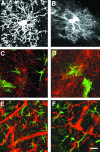
Morphology and distribution of NG2-glia and astrocytes. Confocal microscopic images of mid-sagittal sections of P15 brains from NG2-DsRed mice (A,E,F) and GFAP-EGFP mice (B–D), immunolabelled for NG2 (C,D) or GFAP (E,F). (A,B) NG2-glia and protoplasmic astrocytes are superficially similar, but differ in the organization of their process arborizations and structure. (C,D) Immunolabelled NG2-glia (red) are a separate and distinct population from EGFP+ protoplasmic astrocytes (green). The process domains of NG2-glia and protoplasmic astrocytes overlap with each other. (E,F) GFAP immunolabelling was not observed in DsRed+ NG2 glia; note the DsRed+ blood vessels in E. Scale bar = 25 µm in C,D and 10 µm in A,B,E,F.
Fig. 3
Fig. 3
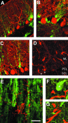
NG2-glia and Bergmann glia have different domains in the cerebellum. Confocal microscopic images of mid-sagittal sections of P19 cerebellum from GFAP-EGFP mice (A,B,C,E) and NG2-DsRed mice (D,F,G), immunolabelled for calbindin (A,B,C,F,G) or NG2 (E). (A–C) EGFP+ Bergmann glia (green) have cell bodies adjacent to calbindin-immunopositive Purkinje cells (red). A single Purkinje cell may be served by multiple Bergmann glia, which extend three or more primary processes from which pass innumerable fine collaterals that contact the dendritic trees of the Purkinje cells. (D) DsRed+ NG2-glia are located throughout the cerebellar layers (some Purkinje cells identified by asterisk), with processes that extend radially through the molecular layer (ML), Purkinje cell layer (PCL) and granule cell layer (GCL). (E) Immunopositive NG2-glial cell in the molecular cell layer extends processes that intertwine with the rising processes of multiple Bergmann glia. (F,G) DsRed+ NG2-glial cells amongst the cell body (F) and dendritic trees (G) of calbindin-immunopositive Purkinje cells. Scale bar = 50 µm in A, 30 µm in B–D, and 20 µm in E–G.
Fig. 4
Fig. 4
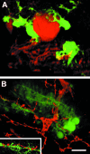
Relations between Bergmann glia, Purkinje neurons and NG2-glia. Confocal microscopic images of mid-sagittal sections of P19 cerebellum from GFAP-EGFP mice, immunolabelled for calbindin (A) or NG2 (B), and deconvolved and rotated using Volocity (version 4.0.1; Improvision, UK). (A) Multiple Bergmann glia serve a single Purkinje cell, and each Bergmann glial cell contains many hundreds of independent microdomains which ensheath synapses. (B) NG2-glia process domains encompasses multiple Bergmann glia and form intricate associations with the processes of Bergmann glia that ensheath synapses (inset). NG2-glia receive inputs via these processes from multiple climbing fibres (Lin et al. 2004). Scale bar = 15 µm.
Fig. 5
Fig. 5
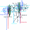
Bergmann glia and NG2-glia are functional elements in the cerebellar network. Bergmann glia (green) and NG2-glia (black), respectively, ensheath and contact synapses formed by parallel fibres (light blue) and climbing fibres (red) on the dendritic tree of Purkinje cells (dark blue) in the molecular layer of the cerebellum. Bergmann glia are orientated parallel to the Purkinje cell dendritic tree, whereas NG2-glia are radially orientated, contacting multiple Purkinje cells and Bergmann glia. Bergmann glia and NG2-glia are anatomically and physiologically integrated into neural networks.
Fig. 6
Fig. 6
Stimulation of axons in the optic nerve evokes calcium signalling in astrocytes and NG2-glia. Optic nerves from GFAP-EGFP mice and NG2-DsRed mice aged P15 were isolated intact and loaded with the calcium-sensitive dye fura-2, as described previously (James & Butt, 2002). Cells were imaged on an Olympus upright microscope (BX50W1), and visualized using an Achroplan ×20 water immersion lens and excited alternately at 340 and 380 nm, using a Cairn monochromator (Cairn Research Ltd, UK). Emissions were detected at 510 nm using a Photometrics S-Coolsnap CCD camera (supplied by Cairn Research Ltd, UK). The monochromator and the CCD camera were controlled and synchronized by Axon Imaging Workbench 5.1 (Axon Instruments), and quantitative measurements were made using the same program. (A) Measurements were made from EGFP+ astrocytes and DsRed+ NG2-glia, and calcium variations expressed as the change in ratio after background subtraction (F340:F380). Spontaneous [Ca2+]i oscillations were observed in both EGFP+ astrocytes and DsRed+ NG2-glia. (B) Electrical stimulation of the optic nerve to evoke axonal action potentials resulted in a rapid increase in [Ca2+]i in EGFP+ astrocytes and DsRed+ NG2-glia, which was sustained for the duration of the stimulus. (C) Bath administration of 100 µm ATP or glutamate evoked raised [Ca2+]i in EGFP+ astrocytes and DsRed+ NG2-glia. (D) Mechanical stimulation of the optic nerve with a sharp microelectrode triggered raised glial [Ca2+]i that was inhibited by the purinoreceptor antagonist suramin, indicating that ATP released by astrocytes propagates a calcium wave that passes to NG2-glia.

Similar articles

Cited by

References

    1. Aguirre A, Chittajallu R, Belachew S, Gallo V. NG2-expressing cells in the subventricular zone are type C-like cells that contribute to interneuron generation in the postnatal hippocampus. J Cell Biol. 2004;165:575–589. - PMC - PubMed
    1. Aguirre A, Gallo V. Postnatal neurogenesis and gliogenesis in the olfactory bulb from NG2-expressing progenitors of the subventricular zone. J Neurosci. 2004;24:10530–10541. - PMC - PubMed
    1. Alberdi E, Sanchez-Gomez MV, Matute C. Calcium and glial cell death. Cell Calcium. 2005;38:417–425. - PubMed
    1. Araque A, Parpura V, Sanzgiri RP, Haydon PG. Tripartite synapses: glia, the unacknowledged partner. Trends Neurosci. 1999;22:208–215. - PubMed
    1. Belachew S, Chittajallu R, Aguirre AA, et al. Postnatal NG2 proteoglycan-expressing progenitor cells are intrinsically multipotent and generate functional neurons. J Cell Biol. 2003;161:169–186. - PMC - PubMed

Publication types

LinkOut - more resources