

Resection of the mesopancreas (RMP): a new surgical classification of a known anatomical space
- PMID: 17459163
- PMCID: PMC1865381
- DOI: 10.1186/1477-7819-5-44
Resection of the mesopancreas (RMP): a new surgical classification of a known anatomical space
Abstract
Background: Prognosis after surgical therapy for pancreatic cancer is poor and has been attributed to early lymph node involvement as well as to a strong tendency of cancer cells to infiltrate into the retropancreatic tissue and to spread along the peripancreatic neural plexuses. The objective of our study was to classify the anatomical-surgical layer of the mesopancreas and to describe the surgical principles relevant for resection of the mesopancreas (RMP). Immunohistochemical investigation of the mesopancreatic-perineural lymphogenic structures was carried out with the purpose of identifying possible routes of metastatic spread.
Methods: Resection of the mesopancreas (RMP) was performed in fresh corpses. Pancreas and mesopancreas were separated from each other and the mesopancreas was immunohistochemically investigated.
Results: The mesopancreas strains itself dorsally of the mesenteric vessels as a whitish-firm, fatty tissue-like layer. Macroscopically, in the dissected en-bloc specimens of pancreas and mesopancreas nerve plexuses were found running from the dorsal site of the pancreatic head to the mesopancreas to establish a perineural plane. Immunohistochemical examinations revealed the lymphatic vessels localized in direct vicinity of the neuronal plexuses between pancreas and mesopancreas.
Conclusion: The mesopancreas as a perineural lymphatic layer located dorsally to the pancreas and reaching beyond the mesenteric vessels has not been classified in the anatomical or surgical literature before. The aim to ensure the greatest possible distance from the retropancreatic lymphatic tissue which drains the carcinomatous focus can be achieved in patients with pancreatic cancer only by complete resection of the mesopancreas (RMP).
Figures
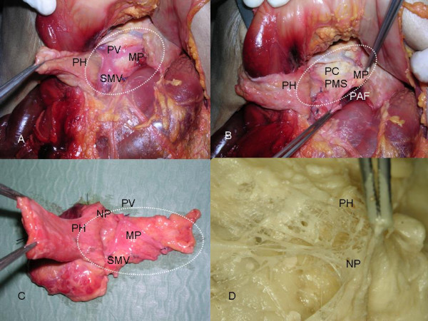
 . B). Resection of the pancreas and mesopancreas dorsally of the mesenteric vessels en bloc. PH = Pancreatic Head; MP = Mesopancreas ; PAF = Preaortic Fascia, PC = Plexus Coeliacus, PMS = Plexus Mesentericus superior. C). Macroscopically, in the dissected en-bloc specimen of the pancreas and mesopancreas nerve plexuses are found running from the dorsal surface of the pancreatic head to the mesopancreas to establish a perineural plane. PH = Pancreatic Head;NP = Nerve Plexus; SMV = Superior Mesenteric Vein; PV = Portal Vein; MP = Mesopancreas . D). The nerve plexus of the dorsal site of the pancreatic head is depicted in a detailed view after preparation of the layer between pancreas and mesopancreas. PH = Pancreatic Head; NP = Nerve Plexus of the dorsal margin of the pancreatic head.
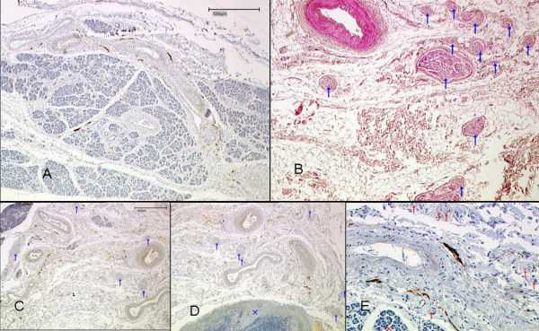
References
-
- Luttges J, Vogel I, Menke M, Henne-Bruns D, Kremer B, Kloppel G. The retroperitoneal resection margin and vessel involvement are important factors determining survival after pancreaticoduodenectomy for ductal adenocarcinoma of the pancreas. Virchows Arch. 1998;433:237–242. doi: 10.1007/s004280050242. - DOI - PubMed
-
- Neoptolemos JP, Stocken DD, Dunn JA, Almond J, Beger H, Pederzoli P. Influence of resection margins on survival for patients with pancreatic cancer treated by adjuvant chemoradiation and/or chemotherapy in the ESPAC-1 randomized controlled trial. Ann Surg. 2001;234:758–768. doi: 10.1097/00000658-200112000-00007. - DOI - PMC - PubMed
MeSH terms
LinkOut - more resources
Full Text Sources
Medical
Research Materials
