. 2007 Apr 25:5:16.
doi: 10.1186/1477-7827-5-16.

Distinct phospholipase A2 enzymes regulate prostaglandin E2 and F2alpha production by bovine endometrial epithelial cells

Affiliations

Distinct phospholipase A2 enzymes regulate prostaglandin E2 and F2alpha production by bovine endometrial epithelial cells

Patricia K Tithof et al. Reprod Biol Endocrinol. .

Abstract

Background: The rate-limiting step in prostaglandin (PG) biosynthesis is catalyzed by phospholipase A2 (PLA2) enzymes which hydrolyze arachidonic acid from membrane phospholipids. Despite their importance in uterine PG production, little is known concerning the specific PLA2 enzymes that regulate arachidonic acid liberation in the uterine endometrium. The objectives of this study were to evaluate the expression and activities of calcium-independent Group VI and Group IVC PLA2 (PLA2G6 and PLA2G4C) and calcium-dependent Group IVA PLA2 (PLA2G4A) enzymes in the regulation of bovine uterine endometrial epithelial cell PG production.

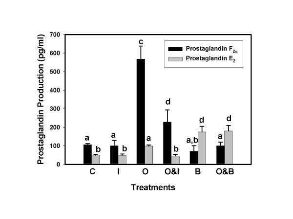
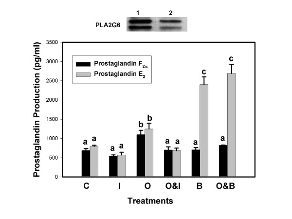
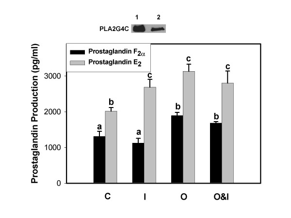
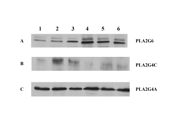
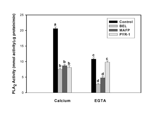
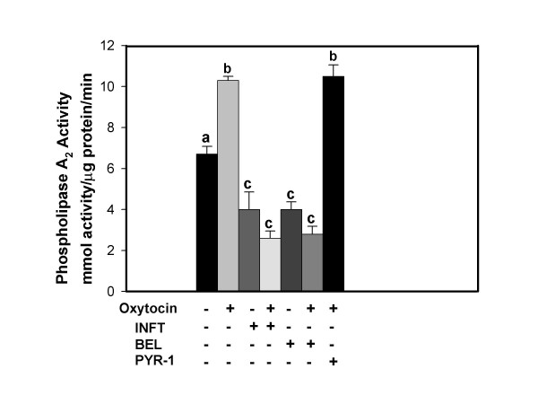
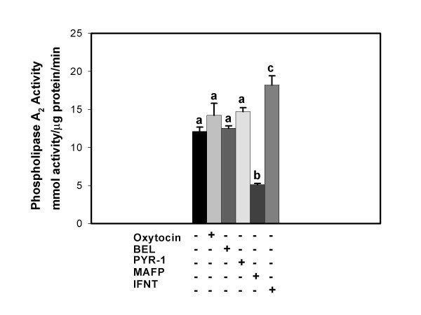
Methods: Bovine endometrial epithelial cells in culture were treated with oxytocin, interferon-tau and the PLA2G6 inhibitor bromoenol lactone, alone and in combination. Concentrations of PGF2alpha and PGE2 released into the medium were analyzed. Western blot analysis was performed on cellular protein to determine the effects of treatments on expression of PLA2G4A, PLA2G6 and PLA2G4C. Group-specific PLA2 activity assays were performed on cell lysates following treatment with oxytocin, interferon-tau or vehicle (control), alone and in combination. To further evaluate the role of specific PLA2 enzymes in uterine cell PG biosynthesis, cells were transfected with cDNAs encoding human PLA2G6 and PLA24C, treated as described above and PG assays performed.

Results: Constitutive cell production of PGF2alpha was about two-fold higher than PGE2. Oxytocin stimulated production of both PGs but the increase of PGF2alpha was significantly greater. Interferon-tau diminished oxytocin stimulation of both PGs. The PLA2G6 inhibitor, bromoenol lactone, abolished oxytocin-stimulated production of PGF2alpha. Treatments had little effect on PLA2G4A protein expression. In contrast, oxytocin enhanced expression of PLA2G6 and this effect was diminished in the presence of interferon-tau. Expression of PLA2G4C was barely detectable in control and oxytocin treated cells but it was enhanced in cells treated with interferon-tau. Oxytocin stimulated PLA2 activity in assays designed to evaluate PLA2G6 activity and interferon-tau inhibited this response. In assays designed to measure PLA2G4C activity, only interferon-tau was stimulatory. Cells overexpressing PLA2G6 produced similar quantities of the two PGs and these values were significantly higher than PG production by non-transfected cells. Oxytocin stimulated production of both PGs and this response was inhibited by interferon-tau. Bromoenol lactone inhibited oxtocin stimulation of PGF2alpha production but stimulated PGE2 production, both in the absence and presence of oxytocin. Cells over-expressing PLA2G4C produced more PGE2 than PGF2alpha and interferon-tau stimulated PGE2 production.

Conclusion: Results from these studies indicate that oxytocin stimulation of uterine PGF2alpha production is mediated, at least in part, by up-regulation of PLA2G6 expression and activity. In addition to its known inhibitory effect on oxytocin receptor expression, interferon-tau represses oxytocin-stimulated PLA2G6 expression and activity and this contributes to diminished PGF2alpha production. Furthermore, endometrial cell PGE2 biosynthesis was associated with PLA2G4C expression and activity and interferon-tau was stimulatory to this process.


Figures

Figure 1
Figure 1
Prostaglandin Fand E2 production by BEE cells. Cell culture medium was harvested 6 h after addition of treatments and concentrations of PGs were analyzed by ELISA. Treatments included, Control, C; IFNT, I; Oxytocin, O; Oxytocin and IFNT, O&I, Bromoenol lactone, B; and Oxytocin and Bromoenol lactone, O&B. Treatments were performed in triplicate and repeated (n = 6). Data are expressed as means and SEM. Columns with different superscripts are significantly different (p < 0.05).
Figure 2
Figure 2
Prostaglandin Fand E2 production by BEE cells over-expressing PLA2G6. Culture medium from BEE cells, transfected with PLA2G6 cDNA, was harvested 6 h after exposure to treatments and concentrations of PGs were analyzed by ELISA. Treatments included Control, C; IFNT, I; Oxytocin, O; :Oxytocin and IFNT, O&I, Bromoenol lactone, B; Oxytocin and Bromoenol lactone, B&O. Treatments were performed in triplicate and repeated (n = 6). Data are expressed as means and SEM. Columns with different superscripts are significantly different (p < 0.05). Western blot at top of figure compares PLA2G6 expression in transfected cells (lane 1) and non-transfected cells (lane 2).
Figure 3
Figure 3
Prostaglandin Fand E2 production by BEE cells over-expressing PLA2G4C. Culture medium from BEE cells, transfected with PLA2G4C cDNA, was harvested 6 h after exposure to treatments and concentrations of PGs were analyzed by ELISA. Treatments included Control, C; IFNT, I; Oxytocin, O, Oxytocin and IFNT, O&I. Data are expressed as means and SEM. Treatments were performed in triplicate and repeated (n = 6). Columns with different superscripts are significantly different (p < 0.05). Western blot at top of figure compares PLA2G4C expression in transfected cells (lane 1) and non-transfected cells (lane 2).
Figure 4
Figure 4
Oxytocin and interferon-tau differentially regulate expression of PLA2 enzymes. BEE cells were treated for 6 h with vehicle (control, lane 1), 50 ng IFNT (lane 2), 1000 ng IFNT (lane 3), oxytocin (lane 4), oxytocin plus 50 ng IFNT (lane 5), and oxytocin plus 1000 ng IFNT (lane 6). Western blot analyses were performed on cellular proteins using antibodies against A, PLA2G6; B, PLA2G4C and C, PLA2G4A as described in the Methods.
Figure 5
Figure 5
BEE cells exhibit both Ca++-dependent and Ca++-independent PLA2 activities. PLA2 activity assays were performed on cellular lysates following treatment of intact cells, as described in the Methods. Ca++-dependent PLA2 activity assays were performed with AA-PC and 14C-AA-PC in the presence of 5 mM CaCl2and Ca++-independent assays were performed using the same substrates but in the absence of CaCL2 and the presence of 5 mM EGTA. Treatments included the PLA2G6 inhibitor BEL, the PLA2G4 and PLA2G6 inhibitor MAFP and the PLA2G4A inhibitor PYR-1. Data from columns with different superscripts are significantly different (p < 0.05).
Figure 6
Figure 6
Oxytocin stimulated PLA2G6 activity is blocked by IFNT. BEE cells were treated for 3 h with vehicle control, oxytocin, IFNT, BEL or PYR-1, alone or in combination. The symbols, + and -, indicate the presence or absence of treatments, respectively. PLA2G6 activity assays were performed on cellular lysates as described in the Methods. Inhibition of activity by BEL but not PYR-1 demonstrates that PLA2G6 activity was measured. Columns with different superscripts are significantly different (p < 0.05).
Figure 7
Figure 7
PLA2G4C activity is enhanced by IFNT but not oxytocin. BEE cells were treated for 3 h with vehicle control, oxytocin, BEL, PYR-1, MAFP or IFNT. The symbols, + and -, indicate the presence or absence of treatments, respectively. PLA2G4C activity assays were performed on cellular lysates as described in the Methods. Inhibition of activity by MAFP but not PYR-1 or BEL demonstrates that PLA2G4C activity was measured. Columns with different superscripts are significantly different (p < 0.05).

Similar articles

Cited by

References

    1. McCracken JA, Custer EE, Lamsa JC. Luteolysis: a neuroendocrine mediated event. Physiol Rev. 1999;79:263–323. - PubMed
    1. Silvia WJ, Lewis GS, McCracken JA, Thatcher WW, Wilson L., Jr Hormonal regulation of uterine secretion of prostaglandin F2α during luteolysis in ruminants. Biol Reprod. 1991;45:655–663. doi: 10.1095/biolreprod45.5.655. - DOI - PubMed
    1. Kindahl H, Lindell JO, Edqvist LE. Release of prostaglandin F2α during the oestrous cycle. Acta Vet Scand. 1981;77:143–158. - PubMed
    1. Schramm W, Bovaird L, Glew ME, Schramm G, McCracken JA. Corpus luteum regression induced by ultra-low pulses of prostaglandin F2α. Prostaglandins. 1983;26:347–364. doi: 10.1016/0090-6980(83)90171-5. - DOI - PubMed
    1. Robert RM. Conceptus interferons and maternal recognition of pregnancy. Biol Reprod. 1989;40:449–452. doi: 10.1095/biolreprod40.3.449. - DOI - PubMed

MeSH terms

LinkOut - more resources