

The opioid receptor antagonist, naloxone, protects spinal motor neurons in a murine model of alphavirus encephalomyelitis
- PMID: 17459376
- PMCID: PMC1939803
- DOI: 10.1016/j.expneurol.2007.03.013
The opioid receptor antagonist, naloxone, protects spinal motor neurons in a murine model of alphavirus encephalomyelitis
Abstract
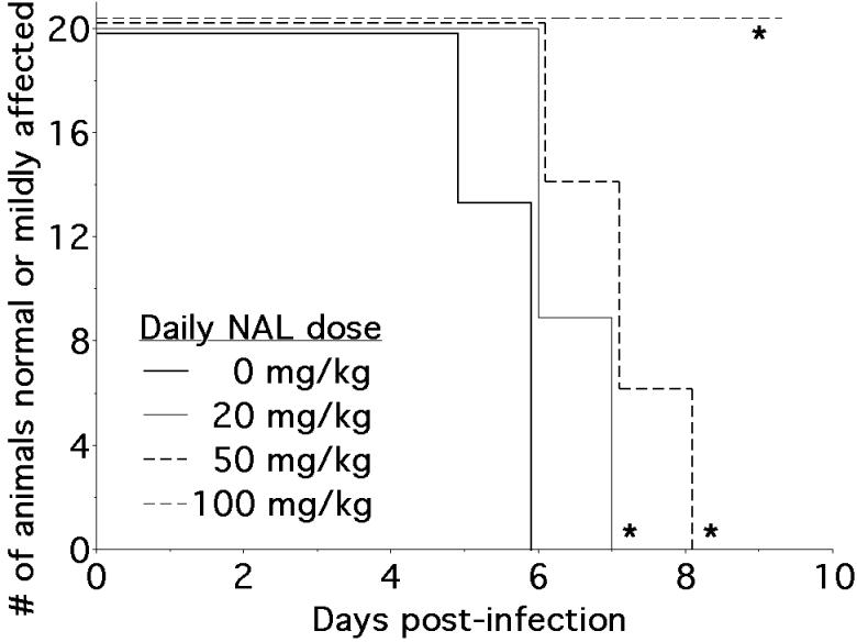
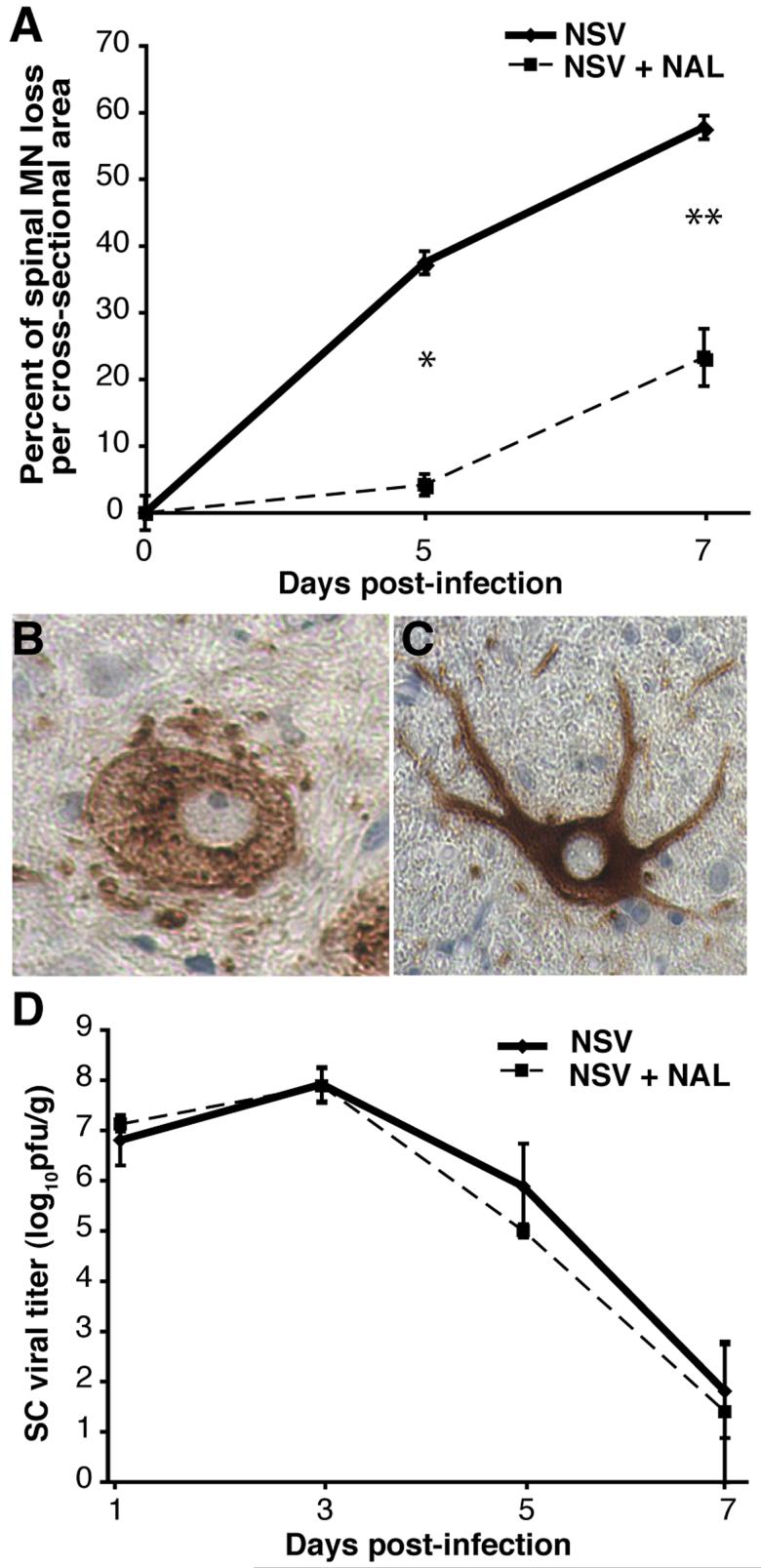
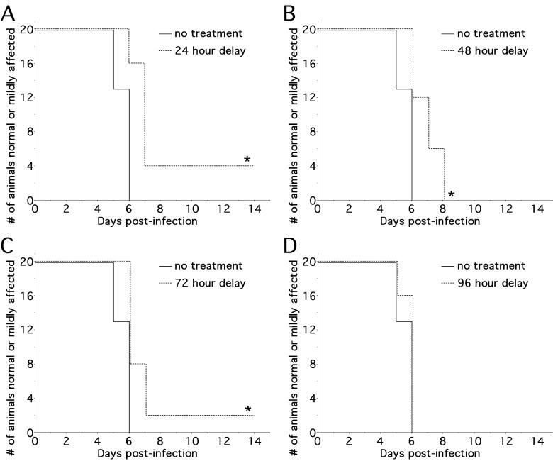
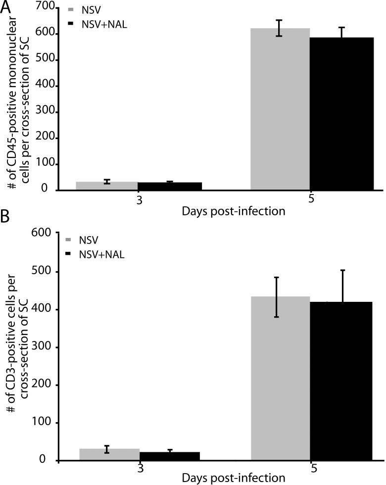
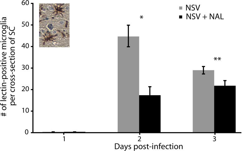
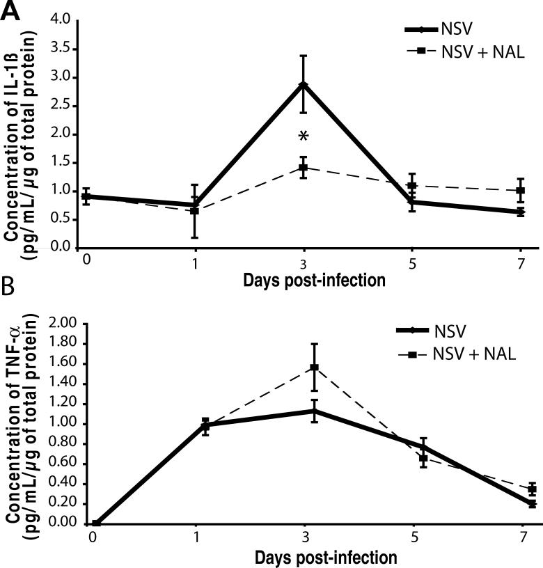
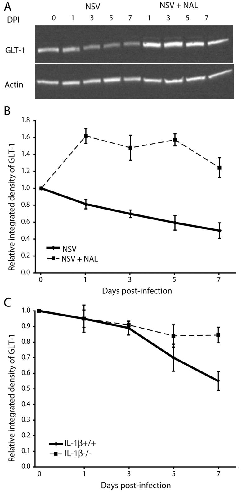
Spread of neuroadapted Sindbis virus (NSV) to motor neurons (MN) of the spinal cord (SC) causes severe hind limb weakness in C57BL/6 mice and models the paralysis that can accompany alphavirus and flavivirus encephalomyelitis in humans. The fate of spinal MN dictates the severity of NSV-induced paralysis, and recent data suggest that MN damage can occur indirectly via the actions of activated microglial cells. Because the opioid receptor antagonist, naloxone (NAL), blocks microglial-mediated neurodegeneration in other models, we examined its effects during NSV infection. Drug treatment prevented paralysis and enhanced the survival of MN without altering NSV tropism, replication, or clearance from SC tissue. Further studies showed that NAL most effectively inhibited paralysis in a 72-h window after NSV challenge, suggesting that the drug inhibits an early event in SC pathogenesis. Histochemical studies demonstrated that NAL blocked early microglial activation in SC tissue sections, and protein assays showed that the early induction of pathogenic IL-1 beta was blunted in SC homogenates. Finally, loss of glutamate transporter-1 (GLT-1) expression in SC, an astrocyte glutamate reuptake protein responsible for lowering toxic extracellular levels of glutamate and preventing MN damage, was reversed by NAL treatment. This GLT-1 loss proved to be highly IL-1 beta-dependent. Taken together, these data suggest that NAL is neuroprotective in the SC by inhibiting microglial activation that, in turn, maintains normal astrocyte glutamate homeostasis. We propose that drugs targeting such microglial responses may have therapeutic benefit in humans with related viral infections.
Figures
Similar articles
-
Viral-induced spinal motor neuron death is non-cell-autonomous and involves glutamate excitotoxicity.J Neurosci. 2004 Aug 25;24(34):7566-75. doi: 10.1523/JNEUROSCI.2002-04.2004. J Neurosci. 2004. PMID: 15329404 Free PMC article.
-
The inflammatory cytokine, interleukin-1 beta, mediates loss of astroglial glutamate transport and drives excitotoxic motor neuron injury in the spinal cord during acute viral encephalomyelitis.J Neurochem. 2008 May;105(4):1276-86. doi: 10.1111/j.1471-4159.2008.05230.x. Epub 2008 Jan 14. J Neurochem. 2008. PMID: 18194440 Free PMC article.
-
Tumor necrosis factor-alpha modulates glutamate transport in the CNS and is a critical determinant of outcome from viral encephalomyelitis.Brain Res. 2009 Mar 31;1263:143-54. doi: 10.1016/j.brainres.2009.01.040. Epub 2009 Feb 3. Brain Res. 2009. PMID: 19368827 Free PMC article.
-
Alphavirus Encephalomyelitis: Mechanisms and Approaches to Prevention of Neuronal Damage.Neurotherapeutics. 2016 Jul;13(3):455-60. doi: 10.1007/s13311-016-0434-6. Neurotherapeutics. 2016. PMID: 27114366 Free PMC article. Review.
-
Neuronal cell death in alphavirus encephalomyelitis.Curr Top Microbiol Immunol. 2005;289:57-77. doi: 10.1007/3-540-27320-4_3. Curr Top Microbiol Immunol. 2005. PMID: 15791951 Review.
Cited by
-
Type-I interferons suppress microglial production of the lymphoid chemokine, CXCL13.Glia. 2014 Sep;62(9):1452-62. doi: 10.1002/glia.22692. Epub 2014 May 14. Glia. 2014. PMID: 24829092 Free PMC article.
-
Protection from fatal viral encephalomyelitis: AMPA receptor antagonists have a direct effect on the inflammatory response to infection.Proc Natl Acad Sci U S A. 2008 Mar 4;105(9):3575-80. doi: 10.1073/pnas.0712390105. Epub 2008 Feb 22. Proc Natl Acad Sci U S A. 2008. PMID: 18296635 Free PMC article.
-
Mechanism of West Nile virus neuroinvasion: a critical appraisal.Viruses. 2014 Jul 18;6(7):2796-825. doi: 10.3390/v6072796. Viruses. 2014. PMID: 25046180 Free PMC article. Review.
-
The Toxoplasma Polymorphic Effector GRA15 Mediates Seizure Induction by Modulating Interleukin-1 Signaling in the Brain.mBio. 2021 Jun 29;12(3):e0133121. doi: 10.1128/mBio.01331-21. Epub 2021 Jun 22. mBio. 2021. PMID: 34154412 Free PMC article.
-
Disrupted glutamate transporter expression in the spinal cord with acute flaccid paralysis caused by West Nile virus infection.J Neuropathol Exp Neurol. 2009 Oct;68(10):1061-72. doi: 10.1097/NEN.0b013e3181b8ba14. J Neuropathol Exp Neurol. 2009. PMID: 19918118 Free PMC article.
References
-
- Acarin L, Vela JM, Gonzalez B, Castellano B. Demonstration of poly-N-acetyl lactosamine residues in ameboid and ramified microglial cells in rat brain by tomato lectin binding. J Histochem Cytochem. 1994;42:1033–1041. - PubMed
-
- Bronstein DM, Perez-Otano I, Sun V, Mullis Sawin SB, Chan J, Wu GC, Hudson PM, Kong LY, Hong JS, McMillian MK. Glia-dependent neurotoxicity and neuroprotection in mesencephalic cultures. Brain Res. 1995;704:112–116. - PubMed
-
- Chang RC, Rota C, Glover RE, Mason RP, Hong JS. A novel effect of an opioid receptor antagonist, naloxone, on the production of reactive oxygen species by microglia: a study by electron paramagnetic resonance spectroscopy. Brain Res. 2000;854:224–229. - PubMed
Publication types
MeSH terms
Substances
Grants and funding
LinkOut - more resources
Full Text Sources
