. 2007 Jul;130(1-2):166-76.
doi: 10.1016/j.pain.2007.03.012. Epub 2007 Apr 24.

Mast cell degranulation activates a pain pathway underlying migraine headache

Affiliations

Mast cell degranulation activates a pain pathway underlying migraine headache

Dan Levy et al. Pain. 2007 Jul.

Abstract

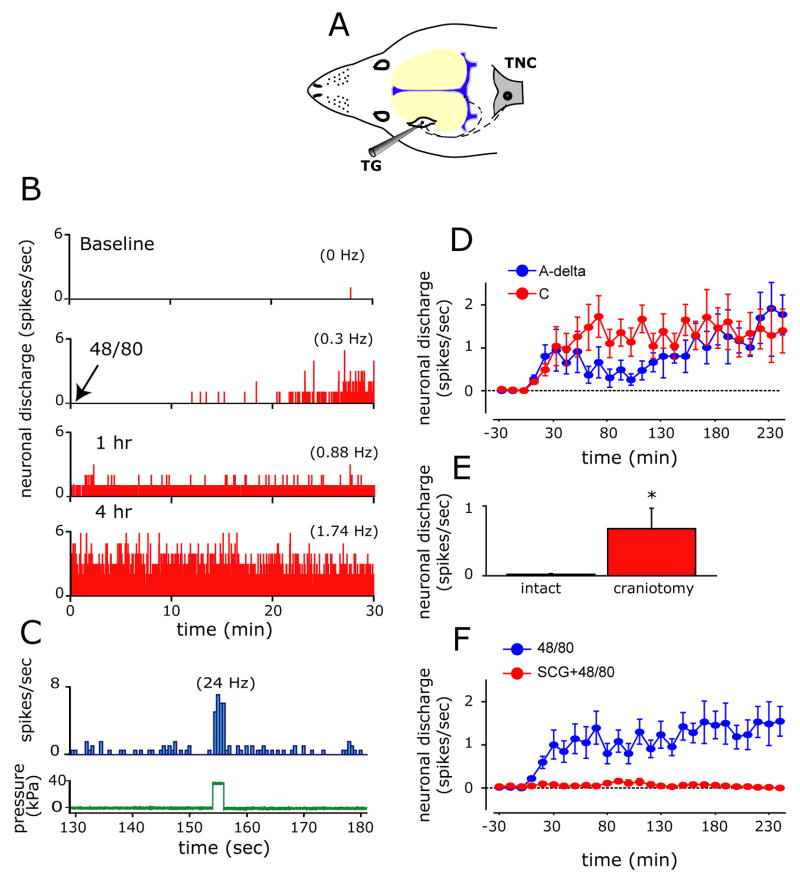
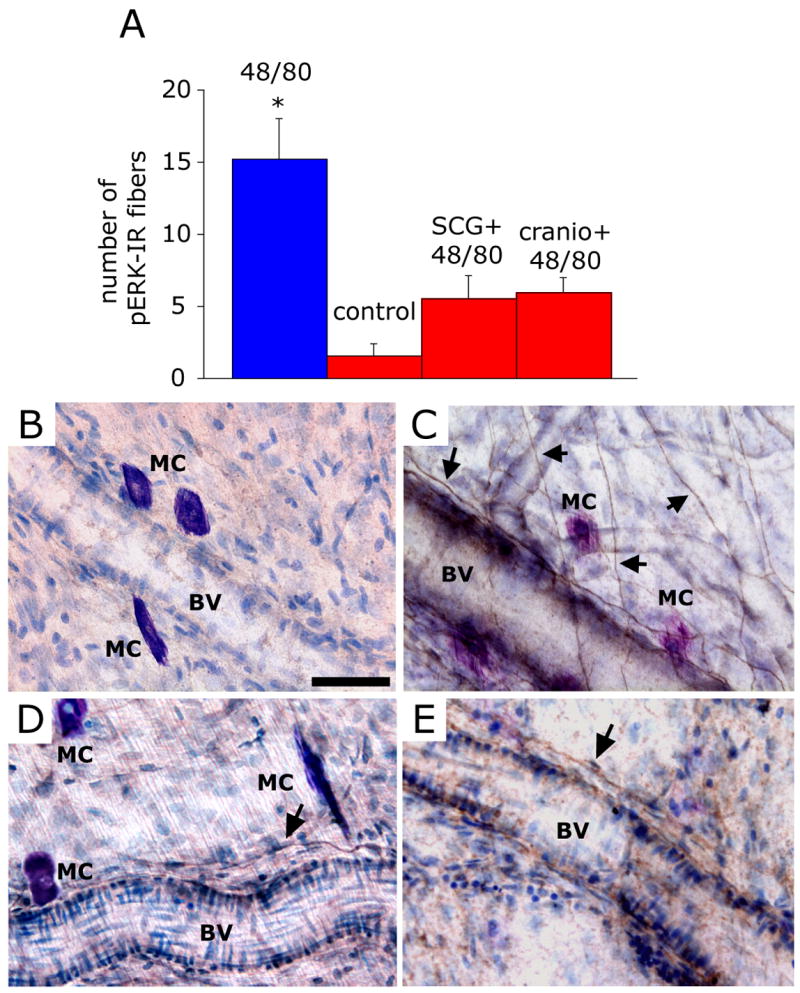
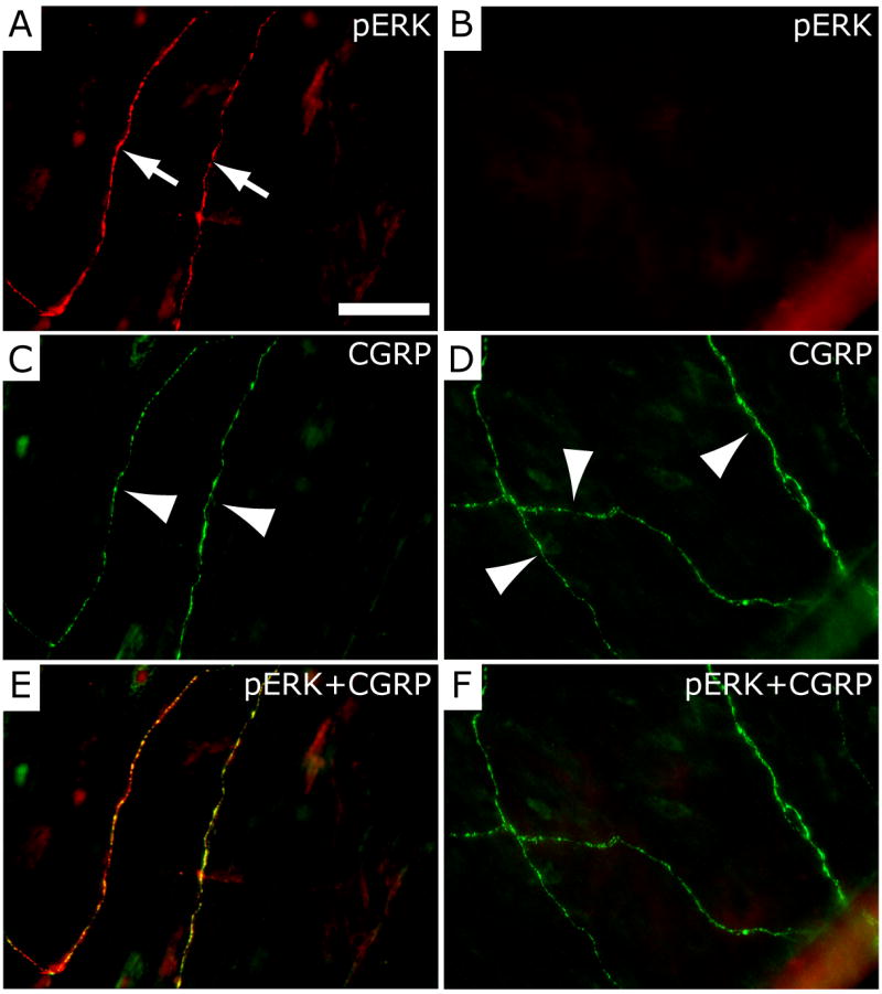
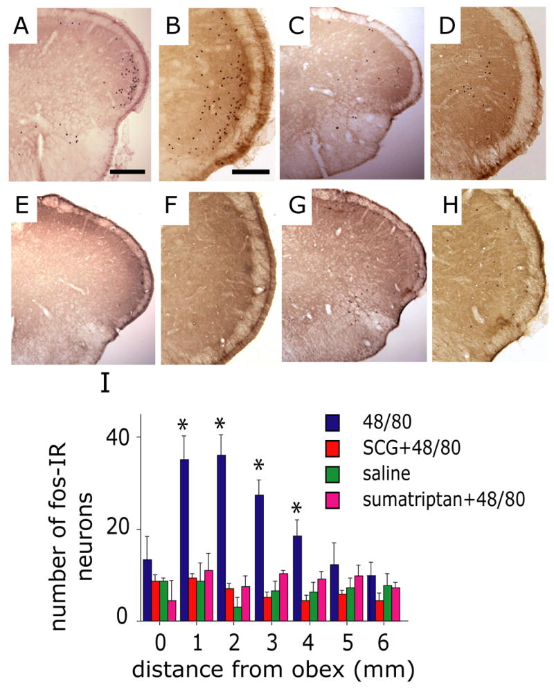
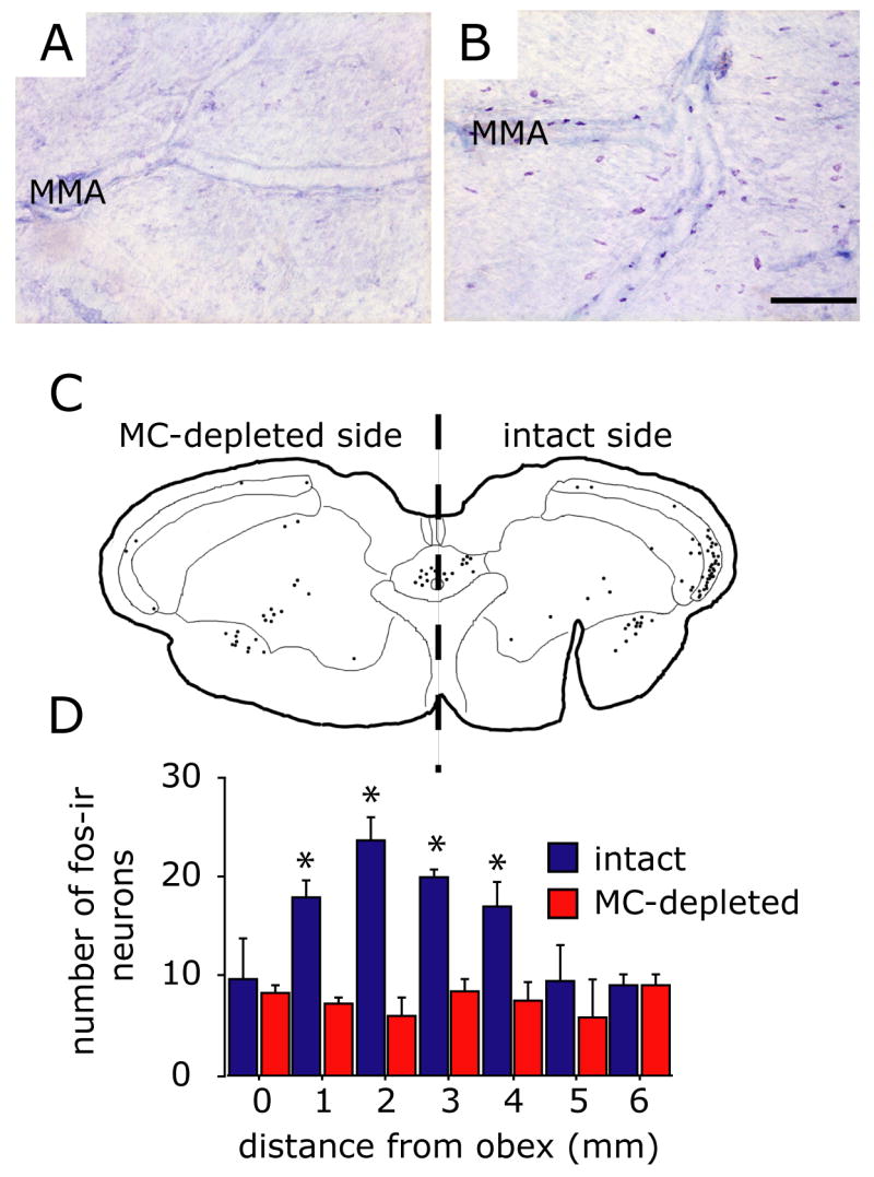
Intracranial headaches such as that of migraine are generally accepted to be mediated by prolonged activation of meningeal nociceptors but the mechanisms responsible for such nociceptor activation are poorly understood. In this study, we examined the hypothesis that meningeal nociceptors can be activated locally through a neuroimmune interaction with resident mast cells, granulated immune cells that densely populate the dura mater. Using in vivo electrophysiological single unit recording of meningeal nociceptors in the rat we observed that degranulation of dural mast cells using intraperitoneal administration of the basic secretagogue agent compound 48/80 (2 mg/kg) induced a prolonged state of excitation in meningeal nociceptors. Such activation was accompanied by increased expression of the phosphorylated form of the extracellular signal-regulated kinase (pERK), an anatomical marker for nociceptor activation. Mast cell-induced nociceptor interaction was also associated with downstream activation of the spinal trigeminal nucleus as indicated by an increase in c-fos expression. Our findings provide evidence linking dural mast cell degranulation to prolonged activation of the trigeminal pain pathway believed to underlie intracranial headaches such as that of migraine.


Figures

Figure 1
Figure 1
Degranulation of dural mast cells by compound 48/80. (A and C) Representative TB-stained dura whole-mount taken from a control animal treated with saline. Note in high magnification (C) the normal appearance of dural MC. (B and D) Toluidine blue - stained dura whole-mount taken 20 minutes following the administration of 48/80. Note the robust signs of degranulation in D. MMA - middle meningeal artery. Scale bar = 500 μm in A and B and 50 μm in C and D.
Figure 2
Figure 2
Mast cell degranulation triggers prolonged excitation of meningeal nociceptors. (A) Schematic localization of the recording site for meningeal nociceptors in the trigeminal ganglion (TG). The figure also illustrates the trigeminovascular pathway from the meninges to the central trigeminal nucleus caudalis (TNC). (B) Selected 30 minutes recording periods demonstrating the neuronal activity (bin size 1 sec) of an affected C-unit meningeal nociceptor at baseline (top) and then at various time points following MC degranulation with 48/80 (bottom traces). Average discharge rates are given in parentheses. (C) Peri-stimulus time histogram showing the response of the same meningeal nociceptor (top trace, bin size 0.5 sec) to a suprathreshold 38kPa mechanical stimulus (bottom trace) applied to the neuron’s dural receptive field following bone removal at the end of the experiment. (D) Time course changes (mean ± SE) in neuronal discharge rate of the affected A-δ and C-units following MC degranulation. (E) The effect of local craniotomy and mast cell depletion on baseline neuronal ongoing discharge level. Note the high level of neuronal activity in neurons in which their receptive field was interrupted by a craniotomy. (F) Time course effect of MC degranulation on meningeal nociceptors showing the increase in ongoing discharge rate (mean ± SEM) and its blockade by prior administration of the MC stabilizer SCG. * p<0.01, Fisher PLSD test.
Figure 3
Figure 3
Mast cell degranulation evokes pERK expression in meningeal nerve fibers. (A) Number (mean ± SEM) of pERK-positive fibers per observation field in the dura of animals treated with 48/80 (n=6), saline (n=4), SCG+48/80 (n=4) and 48/80 (n=4) following local mast cell depletion using craniotomy (cranio + 48/80). * p<0.05 Mann Whitney U test compared with control. (B) Dura whole-mount section taken from control (saline-treated) animal showing non-degranulated MC and lack of pERK expression. (C) An example of typical pERK expression (arrows) in perivascular and non-vascular dural nerve fibers 15 minutes following administration of the MC degranulating agent 48/80. Note the proximity between pERK-positive fibers and the TB-stained degranulated MC. (D) Dura whole-mount section taken from animal treated with SCG prior to 48/80 showing intact MC and very few perivascular pERK-labeled fibers. (E) Dura whole-mount section taken from an animal that underwent a craniotomy prior to 48/80 treatment showing lack of MC staining and very few perivascular pERK-labeled fibers. BV= Blood Vessel. Scale bar = 50 μm
Figure 4
Figure 4
Localization of pERK in CGRP-immunoreactive dural nerve fibers following mast cell degranulation with 48/80. (A) Dural whole-mount section showing pERK immunofluorescence in two dural nerve fibers (arrows) following treatment with 48/80. The same two fibers (arrowheads) showing CGRP-immunofluorescence are shown in C. The merged image (pERK+CGRP) in E shows that the pERK and CGRP labeling is overlapping and thus localized within the same fibers. The dural whole-mount section shown on the right (B, D, F) was obtained from an animal treated with sodium cromoglycate (SCG) prior to 48/80 and shows lack of pERK-immunofluorescence (B), but preservation of CGRP labeling in 3 dural fibers (arrowheads in D). Panel F represents a merged image of B and D. Scale bar = 50 μm.
Figure 5
Figure 5
Mast cell degranulation evokes activation of brainstem trigeminovascular nociceptive neurons. Representative low (A, C, E, G) and high (B, D, F, H) magnification photomicrographs demonstrating c-fos IR in the TNC (medullary dorsal horn) taken from animals treated with 48/80 (A, B), SCG prior to 48/80 (C, D), saline control (E, F) and sumatriptan prior to 48/80 (G, H). Note the distinct distribution of fos in the ventrolateral part of TNC in the 48/80 treated animal. (I) Histogram comparing the number (mean ± SEM) of fos-IR cells in the upper cervical and medullary dorsal horn from animals receiving 48/80, saline, SCG 30 minutes prior to 48/80 or sumatriptan. (* p<0.05, Fisher PLSD test compared with saline control). Scale bar = 1000 μm for (A, C, E, G) and 200 μm for (B, D, F, H).
Figure 6
Figure 6
Depletion of dural mast cell granules blocks 48/80-induced fos expression in the TNC. (A and B) Samples of TB-stained dura mater taken from an animal undergoing a unilateral craniotomy 24 hours earlier. Note that on the craniotomized side (A) there is a complete loss of TB-staining of dural MC indicating granule depletion while the intact, non-craniotomized side (B) shows the typical level of MC degranulation following 48/80 treatment. (C) Cameral Lucida reconstruction of the anatomical location of fos-IR cells within the superficial dorsal horn (laminae I and II) at the level of the caudal TNC following MC degranulation in an animal that underwent a unilateral MC depletion using craniotomy. Each drawn section plots the location of fos-IR cells from 3 consecutive, alternate, 40 mm sections. Note that dural MC depletion blocked fos-IR only in the TNC ipsilateral to the depleted side. (D) Plot of the rostrocaudal distribution of the mean number of fos-IR cells in the dorsal horn ipsi- and contralateral to the MC depleted side in animals injected with 48/80 (n=4). Asterisks indicate a significant difference between the ipsi- and contralateral sides (p<0.05, Fisher PLSD test). Scale bar = 200 μm.

Similar articles

Cited by

References

    1. Artico M, Cavallotti C. Catecholaminergic and acetylcholine esterase containing nerves of cranial and spinal dura mater in humans and rodents. Microsc Res Tech. 2001;53:212–20. - PubMed
    1. Barbara G, Wang B, Stanghellini V, de Giorgio R, Cremon C, Di Nardo G, Trevisani M, Campi B, Geppetti P, Tonini M, Bunnett NW, Grundy D, Corinaldesi R. Mast cell-dependent excitation of visceral-nociceptive sensory neurons in irritable bowel syndrome. Gastroenterology. 2007;132:26–37. - PubMed
    1. Bolay H, Reuter U, Dunn AK, Huang Z, Boas DA, Moskowitz MA. Intrinsic brain activity triggers trigeminal meningeal afferents in a migraine model. Nat Med. 2002;8:136–42. - PubMed
    1. Burstein R, Yamamura H, Malick A, Strassman AM. Chemical stimulation of the intracranial dura induces enhanced responses to facial stimulation in brain stem trigeminal neurons. J Neurophysiol. 1998;79:964–982. - PubMed
    1. Buzzi MG, Moskowitz MA. The antimigraine drug, sumatriptan (GR43175), selectively blocks neurogenic plasma extravasation from blood vessels in dura mater. Br J Pharmacol. 1990;99:202–206. - PMC - PubMed

Publication types

MeSH terms

Substances