. 2007 Jul;81(13):7034-40.
doi: 10.1128/JVI.00029-07. Epub 2007 Apr 25.

Adenovirus E4 34k and E1b 55k oncoproteins target host DNA ligase IV for proteasomal degradation

Affiliations

Adenovirus E4 34k and E1b 55k oncoproteins target host DNA ligase IV for proteasomal degradation

Amy Baker et al. J Virol. 2007 Jul.

Abstract

Cells infected by adenovirus E4 mutants accumulate end-to-end concatemers of the viral genome that are assembled from unit-length viral DNAs by nonhomologous end joining (NHEJ). Genome concatenation can be prevented by expression either of E4 11k (product of E4orf3) or of the complex of E4 34k (product of E4orf6) and E1b 55k. Both E4 11k and the E4 34k/E1b 55k complex prevent concatenation at least in part by inactivation of the host protein Mre11: E4 11k sequesters Mre11 in aggresomes, while the E4 34k/E1b 55k complex participates in a virus-specific E3 ubiquitin ligase that mediates ubiquitination and proteasomal degradation. The E4 34k/E1b 55k complex, but not E4 11k, also inhibits NHEJ activity on internal breaks in the viral genome and on V(D)J recombination substrate plasmids, suggesting that it may interfere with NHEJ independently of its effect on Mre11. We show here that DNA ligase IV, which performs the joining step of NHEJ, is degraded as a consequence of adenovirus infection. Degradation is dependent upon E4 34k and E1b 55k, functional proteasomes, and the activity of cellular cullin 5, a component of the adenoviral ubiquitin ligase. DNA ligase IV also interacts physically with E1b 55k. The data demonstrate that DNA ligase IV, like Mre11, is a substrate for the adenovirus-specific E3 ubiquitin ligase; identify an additional viral approach to prevention of genome concatenation; and provide a mechanism for the general inhibition of NHEJ by adenoviruses.


Figures
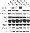
FIG. 1.
FIG. 1.
NHEJ proteins in adenovirus-infected cells. 293 cells were infected with wild-type Ad5 or E4 deletion mutant viruses at a multiplicity of infection of 3.3. Cell lysates were collected 48 h postinfection and fractionated on an SDS-polyacrylamide gel. The gels were analyzed by immunoblotting for E4 34k and proteins of the NHEJ pathway as indicated. The status of open reading frame 6 (ORF 6) (E4 34k) in each mutant is noted on the figure; the complete genotypes of the mutants are listed in Table 1.
FIG. 2.
FIG. 2.
DNA ligase IV in adenovirus-infected cells. (A) 293 cells (left six lanes) or A549 cells (right three lanes) were infected with Ad5 or with E4 or E1b deletion mutant viruses at a multiplicity of infection of 3.3 and harvested 48 h postinfection. (B) 293 cells were transfected with myc-tagged DNA ligase IV and eGFP for 24 h before being infected with Ad5 or E4 deletion mutant viruses and were harvested 48 h postinfection. DNA ligase IV was visualized by immunoblotting with DNA ligase IV antiserum (A) or anti-myc antibody (B). E4 34k was immunoblotted to confirm its expression where expected. Immunoblots of actin (A) or eGFP (B) served as a loading control. The mobility shift of DNA ligase IV in the H5dl1015 lysate is probably a gel artifact due to abundant expression of the viral late protein hexon: alone among viruses which do not degrade DNA ligase IV, H5dl1015 produces high levels of hexon, which migrates immediately above DNA ligase IV on this SDS-polyacrylamide gel. Panel A was assembled from two separate gels.
FIG. 3.
FIG. 3.
DNA ligase IV in E1b 55k-, E4 34k-transfected cells. H1299 cells were transfected with plasmids that express myc-tagged DNA ligase IV (600 ng), E1b 55k and wild-type or mutant (C124S) E4 34k (100 ng each), and eGFP (40 ng). Lysates were harvested 48 h posttransfection and immunoblotted for myc (to detect DNA ligase IV), E4 34k, E1b 55k, cellular actin (loading control; not shown), and eGFP (transfection control). Transfected plasmids are indicated at the top of each lane. The left and right panels are derived from separate experiments. Only lysates of cells transfected with an E4 34k plasmid were probed for E4 34k expression.
FIG. 4.
FIG. 4.
Effect of proteasome inhibition on DNA ligase IV level. 293 cells were mock infected or infected with Ad5 at a multiplicity of infection of 3.3. Infections were carried out in the absence or presence of 10 μM MG132, added 2 h postinfection. Samples were harvested at 12, 24, and 36 h postinfection, and DNA ligase IV was visualized by immunoblotting with DNA ligase IV antiserum. Actin served as a loading control.
FIG. 5.
FIG. 5.
Requirement for cullin 5 in degradation of DNA ligase IV. A549 cells were preinfected with E1-deficient, replication-defective adenovirus vectors expressing FLAG-tagged wild-type (WT) cullin 5 or a dominant-negative carboxy-terminal truncation cullin 5 mutant (NTD) or an empty-vector control. Sixteen hours postinfection, cells were superinfected with Ad5. Fifty-six hours after superinfection, lysates were prepared and analyzed by immunoblotting for expression of cullin 5 (via the FLAG tag), endogenous DNA ligase IV, E4 34k, and cellular actin (loading control). The positions of the full-length and NTD cullin 5 proteins are marked at the left of the bottom panel. The Mock/Mock lysate was not analyzed for either E4 34k or the FLAG tag.
FIG. 6.
FIG. 6.
Physical association of E1b 55k and DNA ligase IV. Lysates were prepared from 293 cells 24 h after cotransfection with plasmids encoding E1b 55k and DNA ligase IV and were incubated with E1b 55k monoclonal antibody 2A6 or an isotype-matched irrelevant control antibody (anti-Ku80). Immune complexes were recovered, fractionated on an SDS-polyacrylamide gel, and analyzed by immunoblotting for DNA ligase IV (top). DNA ligase IV is present in immunoprecipitates made with E1b 55k antiserum (IP 55k) but absent from those made using the irrelevant control (IP Ku), indicating a specific physical interaction between E1b 55k and DNA ligase IV. Immunoprecipitates were also analyzed by immunoblotting with anti-55k serum to confirm successful and specific recovery of E1b 55K (bottom). An amount of anti-55k antibody equal to that used for immunoprecipitation was run in the lane marked “55k Ab” to demonstrate that the band detected by immunoblotting with E1b 55k antiserum is not the result of reactivity of the secondary antibody with immunoglobulin G heavy chain, which is present in the immunoprecipitates and migrates close to E1b 55k in this gel. Lysate equivalent to 15% of the input for an immunoprecipitation was loaded in the first lane.

References

    1. Ahnesorg, P., P. Smith, and S. P. Jackson. 2006. XLF interacts with the XRCC4-DNA ligase IV complex to promote DNA nonhomologous end-joining. Cell 124:301-313. - PubMed
    1. Araujo, F. D., T. H. Stracker, C. T. Carson, D. V. Lee, and M. D. Weitzman. 2005. Adenovirus type 5 E4orf3 protein targets the Mre11 complex to cytoplasmic aggresomes. J. Virol. 79:11382-11391. - PMC - PubMed
    1. Babiss, L. E., and H. S. Ginsberg. 1984. Adenovirus type 5 early region 1b gene product is required for efficient shutoff of host protein synthesis. J. Virol. 50:202-212. - PMC - PubMed
    1. Bender, C. F., M. L. Sikes, R. Sullivan, L. E. Huye, M. M. Le Beau, D. B. Roth, O. K. Mirzoeva, E. M. Oltz, and J. H. Petrini. 2002. Cancer predisposition and hematopoietic failure in Rad50(S/S) mice. Genes Dev. 16:2237-2251. - PMC - PubMed
    1. Boehmer, P. E., and I. R. Lehman. 1997. Herpes simplex virus DNA replication. Annu. Rev. Biochem. 66:347-384. - PubMed

Publication types

MeSH terms