. 2007 Jul;81(13):7149-55.
doi: 10.1128/JVI.00215-07. Epub 2007 Apr 25.

Atypical bZIP domain of viral transcription factor contributes to stability of dimer formation and transcriptional function

Affiliations

Atypical bZIP domain of viral transcription factor contributes to stability of dimer formation and transcriptional function

Celine Schelcher et al. J Virol. 2007 Jul.

Abstract

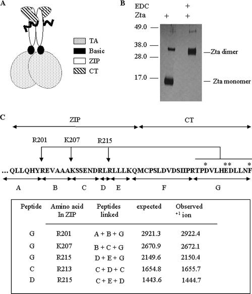
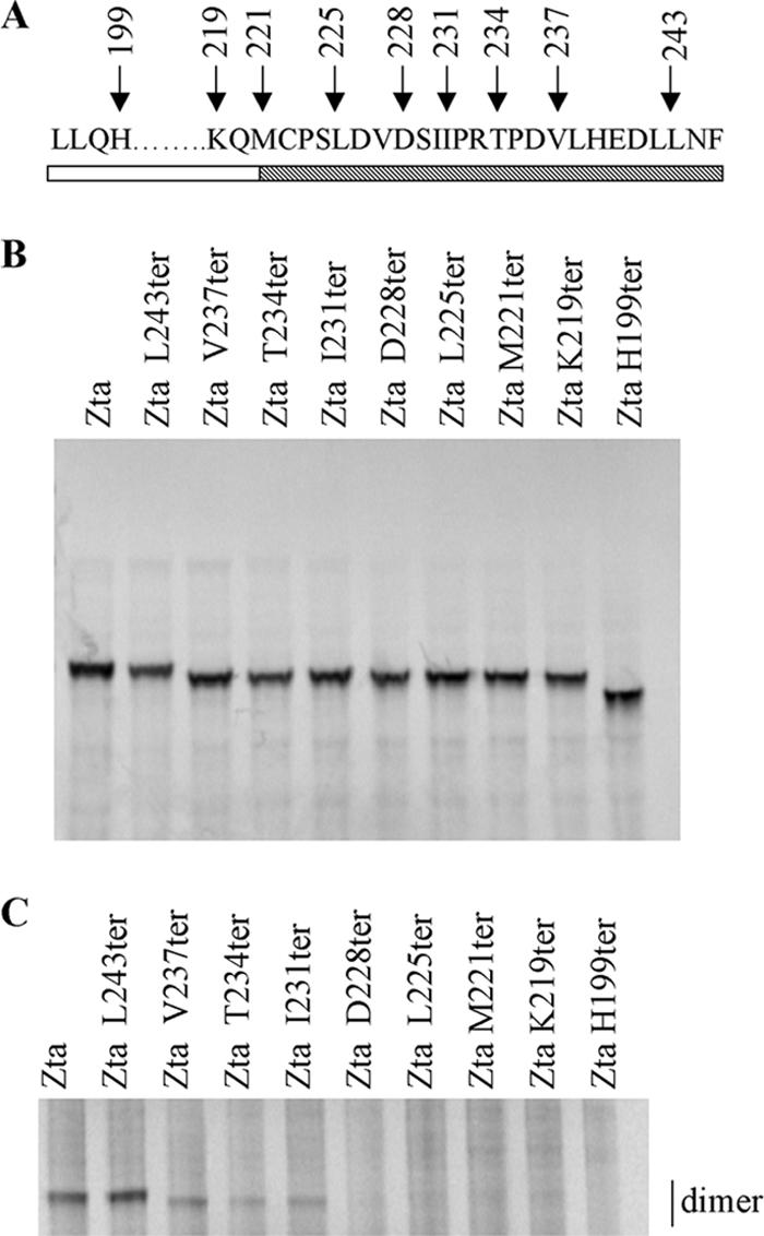
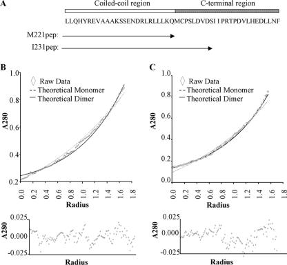
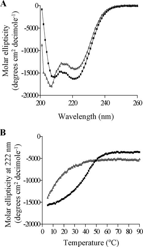
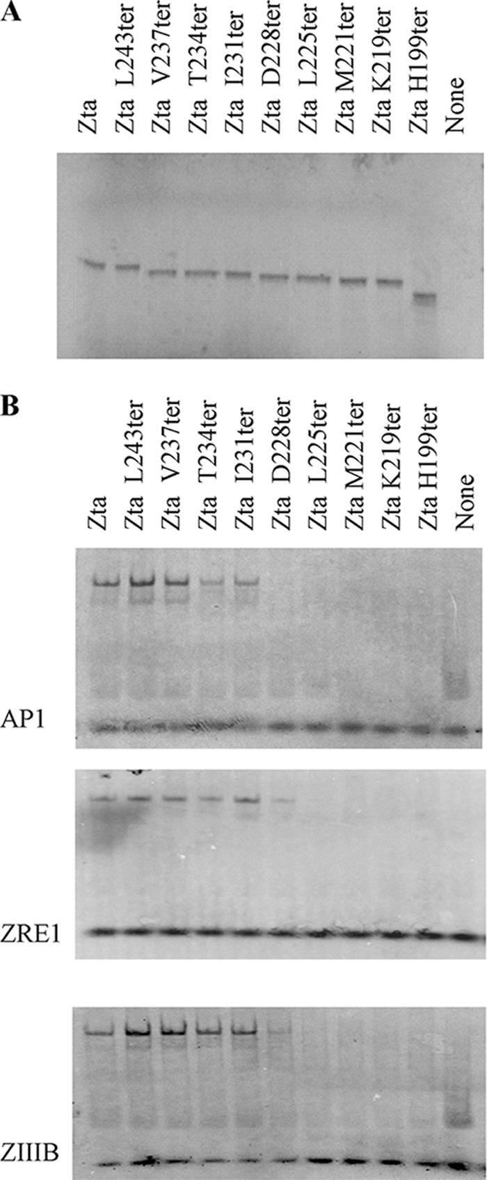
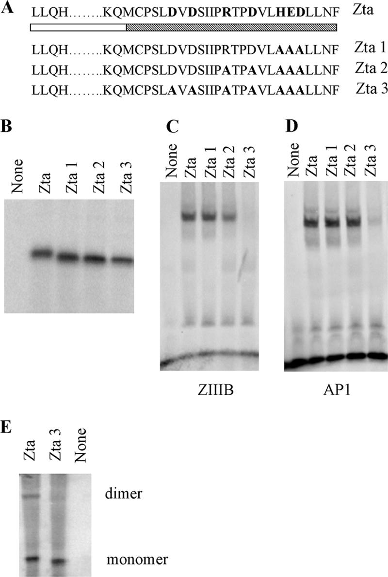
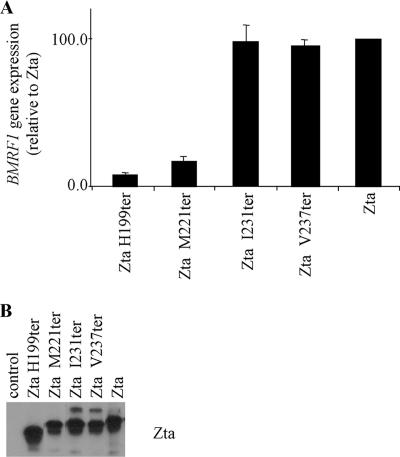
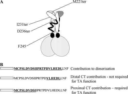
The Epstein-Barr virus transcription factor Zta (encoded by BZLF1) is a bZIP protein containing an alpha-helical coiled-coil homodimerization motif (zipper). The Zta zipper forms less-stable dimers than other bZIP proteins, and an adjacent region (CT) interacts with the zipper to form a novel structure that is proposed to strengthen the dimer. Here we question the role of the CT region for Zta function. Cross-linking experiments demonstrate that the entire CT region lies adjacent to the zipper. Detailed analyses of Zta truncation mutations identify an involvement of the proximal CT region (221 to 230) in dimer formation with a further contribution from the distal region (236 to 243). Biophysical analyses reveal that residues 221 to 230 enhance the stability of the coiled coil. The ability of the Zta truncation mutants to interact with three Zta-binding sites also requires the proximal CT region. Fine mapping of DNA-binding requirements highlighted the contribution of these amino acids for Zta function. Thus, the proximal part of the CT region is required to aid the dimerization of Zta and thereby its DNA-binding ability. In contrast, although the distal part of the CT region aids dimerization, it promotes only a modest increase in DNA binding. To probe this further, we defined the contribution from the CT region for Zta to transactivate a promoter embedded within the viral genome. From this we conclude that the proximal part of the CT region is absolutely required, whereas the distal part is dispensable.


Figures

FIG. 1.
FIG. 1.
The distal CT region closely interacts with the zipper region of Zta. (A) Schematic diagram of Zta showing interaction of part of the CT region with the zipper as determined by Petosa and colleagues (23). The wavy line represents the part of the CT region of unknown structure. (B) Migration of the bZIPCT protein in SDS-PAGE before and after cross-linking with EDC. (C) Following excision and trypsin digestion, resulting peptides were identified by mass spectrometry. Potential tryptic peptides are indicated beneath the sequence, and asterisks indicate acidic residues in peptide G. Peptides A, B, and D cross-linked to the extreme CT region (peptide G) and peptides C and D linked to the zipper were identified, allowing identification of specific cross-linked residues in the ZIP region.
FIG. 2.
FIG. 2.
Both the proximal and distal parts of the CT region contribute to dimerization. (A) Schematic diagram showing sequence of the truncation mutants spanning the zipper and CT regions. The zipper region is shown as an open box and the CT region as a cross-hatched box. (B) Amounts of translation product generated from wheat germ extract for Zta and Zta truncated mutants used in the dimerization assay were assessed by SDS-PAGE and quantitated by phosphorimaging. (C) Equivalent amounts of each protein were incubated at 37°C and cross-linked by addition of 0.05% glutaraldehyde for 30 min. The reaction was quenched by the addition of glycine, and dimeric proteins were separated on a 10% bis-Tris gel and visualized using phosphorimaging. A representative gel is shown.
FIG. 3.
FIG. 3.
Stability of dimers revealed by analytical ultracentrifugation. (A) Schematic representation of residues contained within the two synthetic peptides and their locations compared to Zta structure. Representative analytical ultracentrifuge data sets for M221pep (B) and I231pep (C), recorded at 50,000 rpm, are shown and provide curves typical of sedimented and equilibrated species. Protein concentration (measured as A280) versus distance (r) from the center of the centrifuge rotor. The starting protein concentration was 500 μM. Experimental data points are shown for sedimentation of the peptide (diamonds). Also shown are simulated curves calculated assuming a completely monomeric (broken line) or dimeric (solid line) protein. The residual signals, calculated as the difference between the experimental and fitted data for a single ideal species, show no systematic errors, indicating that the fits are robust (lower panels).
FIG. 4.
FIG. 4.
CD spectroscopy reveals that the C-terminal region of Zta contributes to stability of the coiled-coil dimer. (A) CD analysis determining the secondary structure of the M221pep and I231pep (at a concentration of 100 mM), shown by white triangles and black squares, respectively. (B) The melting curve was obtained by measuring a signal obtained at the wavelength of 222 nm for M221pep or I231pep through a range of temperature (from 2°C to 90°C).
FIG. 5.
FIG. 5.
Requirement of the proximal CT region for interaction with three ZREs. (A) Proteins were assessed by SOS-PAGE. Abilities of indicated Zta truncation mutants to bind to ZREs were determined by EMSA analysis. In all cases the probe was in excess. Following separation of free and bound ZREs by electrophoresis, locations of the complexes were determined using phosphorimaging, with subsequent quantitation. Duplicate experiments were undertaken, and representative images are shown (B).
FIG. 6.
FIG. 6.
Importance of residues 226 and 228 for DNA-binding function of Zta. (A) Schematic representation of mutations of Zta1, Zta2, and Zta3, with the shading as described in Fig. 2. (B) Amounts of translation product generated for Zta and Zta truncated mutants used in the dimerization assay were assessed by SDS-PAGE and quantitated by phosphorimaging. (C and D) Equivalent amounts of protein were analyzed for their ability to bind DNA by EMSA analysis. Following separation of free and bound ZREs by electrophoresis, the locations of the complexes were detected using phosphorimaging, with subsequent quantitation. Duplicate experiments were undertaken, and representative images are shown for ZIIIB (C) and AP1 (D). (E) Equivalent amounts of protein were assessed for their abilities to form dimers as described in the legend to Fig. 2. Proteins of both monomer and dimer sizes are shown.
FIG. 7.
FIG. 7.
The CT region is required for transactivation in vivo. (A) Expression vectors for indicated Zta truncation mutants were introduced into 293-BZLF1-KO cells, and expression of a viral gene, embedded in the genome, was detected using quantitative real-time PCR. Expression of Zta RNA was also detected and was used as a measure of transfection efficiency. The level of expression of the housekeeping gene, L32, was used to standardize signals. Experiments were undertaken in duplicate, and relative expression of BMRF1 was determined. These levels were then expressed relative to the level seen following expression of Zta. (B) Total protein extracts from cells were analyzed for expression of Zta using Western blot analysis.
FIG. 8.
FIG. 8.
Schematic diagram of structure of Zta and requirement of the CT region for function. (A) The schematic diagram of the structure of Zta is based on the crystal structure of Zta, which ends at D236 (23). The additional information that the distal part of the CT region (ending at F245) contacts the zipper and locations of the termination mutants I231ter and M221 ter are shown. (B) Locations of amino acids that contribute to dimerization are shown in bold and underlined. Each line represents mutants that support the conclusion shown on the right-hand side. Transactivation in vivo is represented as TA.

References

    1. Al Mehairi, S., E. Cerasoli, and A. J. Sinclair. 2005. Investigation of the multimerization region of the Kaposi's sarcoma-associated herpesvirus (human herpesvirus 8) protein K-bZIP: the proposed leucine zipper region encodes a multimerization domain with an unusual structure. J. Virol. 79:7905-7910. - PMC - PubMed
    1. Bhende, P. M., W. T. Seaman, H. J. Delecluse, and S. C. Kenney. 2004. The EBV lytic switch protein, Z, preferentially binds to and activates the methylated viral genome. Nat. Genet. 36:1099-1104. - PubMed
    1. Cayrol, C., and E. Flemington. 1996. G(0)/G(1), growth arrest mediated by a region encompassing the basic leucine zipper (bZIP) domain of the Epstein-Barr virus transactivator Zta. J. Biol. Chem. 271:31799-31802. - PubMed
    1. Cayrol, C., and E. K. Flemington. 1996. The Epstein-Barr virus bZIP transcription factor Zta causes G(0)/G(1) cell cycle arrest through induction of cyclin-dependent kinase inhibitors. EMBO J. 15:2748-2759. - PMC - PubMed
    1. Chang, Y. N., D. L. Y. Dong, G. S. Hayward, and S. D. Hayward. 1990. The Epstein-Barr-Virus Zta transactivator—a member of the bZip family with unique DNA-binding specificity and a dimerization domain that lacks the characteristic heptad leucine zipper motif. J. Virol. 64:3358-3369. - PMC - PubMed

Publication types

MeSH terms

LinkOut - more resources