. 2007 Jul;81(13):7220-9.
doi: 10.1128/JVI.00137-07. Epub 2007 Apr 25.

Viral alteration of cellular translational machinery increases defective ribosomal products

Affiliations

Viral alteration of cellular translational machinery increases defective ribosomal products

Peter Berglund et al. J Virol. 2007 Jul.

Abstract

Here we show that cells expressing genes inserted into Semliki Forest virus (SFV) vectors generate a large fraction of defective ribosomal products (DRiPs) due to frequent initiation on downstream Met residues. In monopolizing the host cell translational machinery, SFV reduces levels of translation eukaryotic initiation factor 4E (eIF4E), diminishes phosphorylation of ribosome subunit S6, and phosphorylates translation initiation factor eIF2alpha. We show that the last event is required for SFV mistranslation of inserted genes. Downstream initiation is suppressed by fusing inserted genes with the open reading frame encoding the SFV capsid, demonstrating that one function of the capsid element is to enable ribosomes to initiate translation in the proper location. These results show that in modifying translation, viral vectors can unpredictably increase the generation of truncated polypeptides and thereby the DRiP fraction of inserted gene products, which can potentially affect their yield, therapeutic efficacy, and immunogenicity.


Figures

FIG. 1.
FIG. 1.
NP localizes partially to mitochondria when expressed by an SFV vector. Confocal microscopy was used to study the subcellular localization of NP. HeLa cells were infected for 7 h with SFV-NP (A and B) or SFVC-NP (C) and fixed and processed for indirect immunofluorescence staining using the MAb IC5-3A8 specific for folded NP (α-F), human α-mitochondria auto-Abs (α-mt), or rabbit sera raised against either the NP COOH terminus (α-C) (panels A and C) or the NH2 terminus (α-N) (panel B). (A inset) Cryoelectron microscopy image of a mitochondrion in a HeLa cell infected with SFV-NP and stained with gold-conjugated α-rabbit immunoglobulin Abs to identify binding of α-C Abs.
FIG. 2.
FIG. 2.
A significant fraction of NP expressed from SFV vector is synthesized as truncated forms. BHK cells were infected for 7 h with SFV-NP, SFVC-NP, IAV (Flu), or mock control, as indicated. Cells were metabolically labeled with [35S]Met for 5 min and NP-40 lysates were incubated three times sequentially with beads loaded with IC5-3A8 specific for folded NP (I, II, and III; lanes F) followed by beads loaded with α-COOH pAbs (IV; lanes C). Samples were analyzed by SDS-PAGE followed by quantitative autoradiography. Numbers beside the upper bands indicate the percentage fractions of NP that reacts with the anti-COOH pAbs (IP IV) out of all full-length NP (sums of IPs I to IV, upper area). Numbers beside the lower regions indicate the fractions of shorter fragments reactive to the anti-COOH antiserum (IP IV, lower area) as percentages of all material immunoprecipitated with this antiserum (IP IV, sum of lower and upper regions).
FIG. 3.
FIG. 3.
NH2-terminal truncated NP localizes to mitochondria. (a) BHK cells were transfected with plasmid DNA encoding truncated NP fragments initiating at the Met residue indicated, corresponding to the first through seventh AUGs in the NP sequence. Total cell lysates were subjected to SDS-PAGE, and NP fragments were visualized by immunoblotting with α-COOH pAbs. aa, amino acid. Predicted sizes are given in kilodaltons. (b) Immunofluorescence analysis of cells transfected with indicated NP variant constructs (1 to 7) or infected with SFV-NP or SFVΔAUG-NP. NP was identified using α-COOH pAbs (green) and mitochondria with human auto-Abs (red).
FIG. 4.
FIG. 4.
NP is not translated from truncated mRNA in the context of SFV expression vectors. (Top) Design of primer sets used for the amplification of upstream and downstream regions of the NP gene; (bottom) quantitative real-time PCR analysis of cDNA reverse transcribed from RNA isolated from cells infected with IAV (Flu), SFV-NP, or SFVC-NP and analyzed by three primer sets as indicated. A series of standards was used to determine the absolute concentrations of DNA detected. The amount of cDNA corresponding to amplicons 107 to 229 and 244 to 345 were not lower than the amounts measured for amplicons 974 to 1084. Similar results were obtained in three independent experiments.
FIG. 5.
FIG. 5.
Downstream translation initiation by SFV vector extends to other gene products. Cells were infected with SFV (SFV HA) or vaccinia virus (Unt vac HA) vectors encoding IAV HA. HA species present in cell lysates were resolved using SDS-PAGE and identified by immunoblotting using MAbs specific for the unfolded HA2 domain of HA. α-Actin Abs were also used after stripping of immunoblots to demonstrate the loading of equal amounts cellular material (bottom). (Top) Positions of AUG codons (Met) and putative molecular masses for translation products generated by initiation at those positions. Note that the main portion of full-length HA is glycosylated and migrates with an apparent size greater than 63.4 kDa. Interestingly, the sequences flanking the second, third, and fourth AUG codons were AGGATGA, TCAATGC, and AGGATGG, respectively, with the latter being the most optimal Kozak ribosome binding site.
FIG. 6.
FIG. 6.
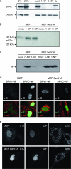
Translational initiation from downstream AUG codons requires phosphorylation of eIF2α. (a) SFV induces reduction in phospho-eIF4E levels. BHK-21 cells were infected with SFV-NP, SFVC-NP, or IAV (flu) as indicated. As controls, cells were either incubated in medium lacking bovine serum (0%) or incubated in medium containing elevated levels (20%). Lysates harvested at 12 h p.i. were subjected to SDS-PAGE and processed for Western blotting using antiserum specific for phospho-eIF4E or actin. (b) SFV induces phosphorylation of eIF2α. MEF obtained either from WT mice or from mice expressing a mutant variant of eIF2α unable to become phosphorylated (Ser51A) were infected with SFV-NP or SFVC-NP as indicated. Lysates harvested at 12 h p.i. were subjected to SDS-PAGE and processed for Western blotting using antiserum specific for phospho-Ser51-eIF2α. (c) Both virus-induced protein synthesis inhibition and the ability of the SFV capsid gene to serve as a translation enhancer require phosphorylation of eIF2α. Lysates from WT cells (MEF) and mutant cells unable to phosphorylatable eIF2α (MEF Ser51A) were infected with SFV-NP or SFVC-NP and analyzed by Western blotting for the expression of NP. The expression from the SFV vector does not suffer from host cell translational shutdown in mutant cells (rightmost lane). (d) Mitochondrial targeting of SFV-expressed NP requires phosphorylation of eIF2α. WT (MEF) and mutant cells unable to phosphorylate eIF2α (MEF Ser51A) were infected with SFV-NP and SFVC-NP as indicated and processed for immunofluorescence using Abs to the carboxy-terminal NP epitope (α-C), fully folded NP (α-F), or mitochondria (α-mt). Confocal images were obtained using identical settings for exposure and laser intensity between the samples illustrating the various expression levels. Mitochondrial NP was detectable only in WT cells infected with SFV-NP (upper left image). (e) Mitochondrial targeting of SFV1-NP requires phosphorylation of eIF2α. Immunolabeled samples of WT (MEF) and mutant cells (MEF Ser51A) from the same immunolabeled SFV1-NP specimens shown in Fig. 6d, with the exception that the settings during the confocal imaging were adjusted in order to acquire next-to-saturated images, thus illustrating the mitochondrial localization of SFV1-NP in WT cells stained with the α-C pAb.
FIG. 7.
FIG. 7.
eIF2α phosphorylation-dependent changes in antigen presentation efficiency. WT and Ser51 MEF were infected with SFV-NP-S-EGFP. Quantitative cytofluorography was used to measure transgene expression as determined by EGFP autofluorescence (left). Antigen presentation was measured simultaneously using Alexa Fluor 647-25-D1.16 (right). Specific presentation was computed as arbitrary units dividing the mean fluorescence intensity levels (background subtracted) corresponding to the 25.D staining with the EGFP fluorescence. (Bottom) The experiment was repeated four additional times with similar results, as indicated in the table.

References

    1. Berglund, P., M. N. Fleeton, C. Smerdou, and P. Liljestrom. 1999. Immunization with recombinant Semliki Forest virus induces protection against influenza challenge in mice. Vaccine 17:497-507. - PubMed
    1. Berglund, P., M. Sjoberg, H. Garoff, G. J. Atkins, B. J. Sheahan, and P. Liljestrom. 1993. Semliki Forest virus expression system: production of conditionally infectious recombinant particles. Bio/Technology 11:916-920. - PubMed
    1. Berglund, P., C. Smerdou, M. N. Fleeton, I. Tubulekas, and P. Liljestrom. 1998. Enhancing immune responses using suicidal DNA vaccines. Nat. Biotechnol. 16:562-565. - PubMed
    1. Bushell, M., and P. Sarnow. 2002. Hijacking the translation apparatus by RNA viruses. J. Cell Biol. 158:395-399. - PMC - PubMed
    1. Davis, N. L., K. W. Brown, and R. E. Johnston. 1996. A viral vaccine vector that expresses foreign genes in lymph nodes and protects against mucosal challenge. J. Virol. 70:3781-3787. - PMC - PubMed

Publication types

MeSH terms

Substances

LinkOut - more resources