. 2007 May 1;104(18):7534-9.
doi: 10.1073/pnas.0702279104. Epub 2007 Apr 25.

Nitric oxide mediates prostaglandins' deleterious effect on lipopolysaccharide-triggered murine fetal resorption

Affiliations

Nitric oxide mediates prostaglandins' deleterious effect on lipopolysaccharide-triggered murine fetal resorption

J Aisemberg et al. Proc Natl Acad Sci U S A. .

Abstract

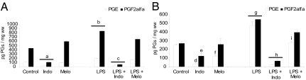
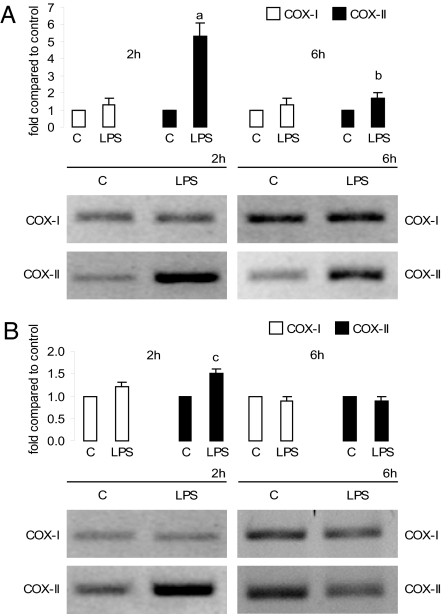
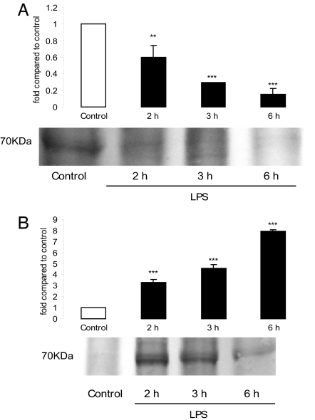
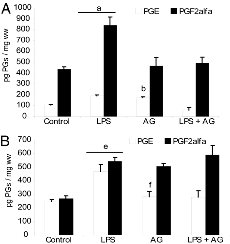
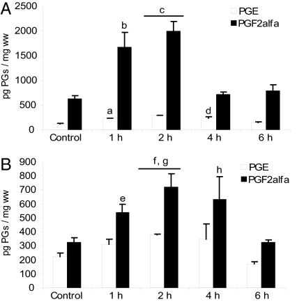
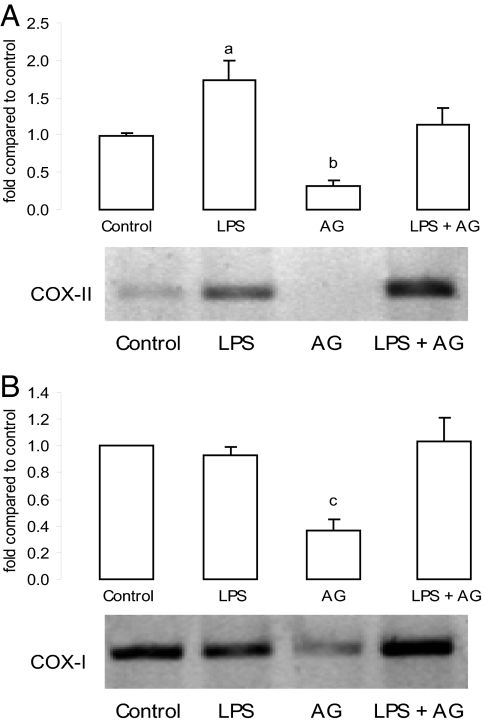
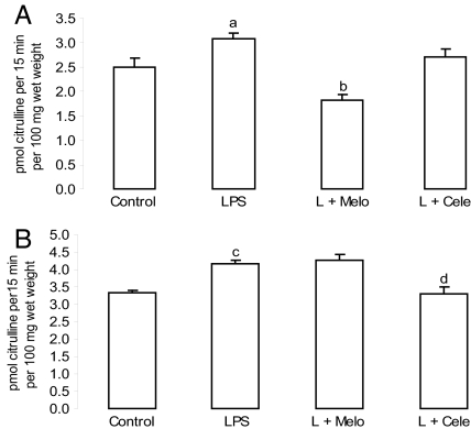
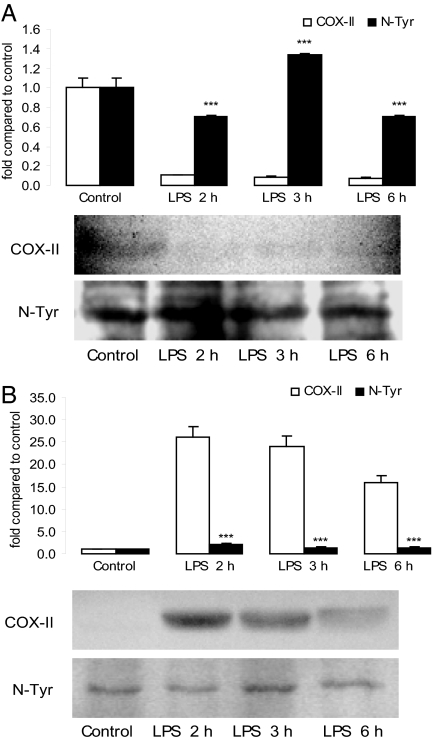
Genital tract bacterial infections could induce abortion and are some of the most common complications of pregnancy; however, the mechanisms remain unclear. We investigated the role of prostaglandins (PGs) in the mechanism of bacterial lipopolysaccharide (LPS)-induced pregnancy loss in a mouse model, and we hypothesized that PGs might play a central role in this action. LPS increased PG production in the uterus and decidua from early pregnant mice and stimulated cyclooxygenase (COX)-II mRNA and protein expression in the decidua but not in the uterus. We also observed that COX inhibitors prevented embryonic resorption (ER). To study the possible interaction between nitric oxide (NO) and PGs, we administered aminoguanidine, an inducible NO synthase inhibitor. NO inhibited basal PGE and PGF(2alpha) production in the decidua but activated their uterine synthesis and COX-II mRNA expression under septic conditions. A NO donor (S-nitroso-N-acetylpenicillamine) produced 100% ER and increased PG levels in the uterus and decidua. LPS-stimulated protein nitration was higher in the uterus than in the decidua. Quercetin, a peroxynitrite scavenger, did not reverse LPS-induced ER. Our results suggest that in a model of septic abortion characterized by increased PG levels, NO might nitrate and thus inhibit COX catalytic activity. ER prevention by COX inhibitors adds a possible clinical application to early pregnancy complications due to infections.


Conflict of interest statement

The authors declare no conflict of interest.

Figures

Fig. 1.
Fig. 1.
In vivo effect of LPS and COX inhibitors on PG production. (A) Uterus. (B) Decidua. Day-7 pregnant mice were injected with PBS (control), LPS (1 μg/g), LPS + Melo (4 mg/kg), or LPS + Indo (2.5 mg/kg) and were killed 6 h after LPS treatment. Values are means ± SEM (n = 8). a and b, P < 0.001 vs. control group; c, P < 0.001 vs. LPS-treated mice; d, P < 0.001 vs. control group; e and f, P < 0.05 vs. control group; g, P < 0.001 vs. control group; h, P < 0.001 vs. LPS-treated mice; and i, P < 0.05 vs. LPS-treated mice.
Fig. 2.
Fig. 2.
In vivo effect of LPS on COX mRNA levels. Uterus (A) and decidua (B) after LPS (1 μg/g) or PBS [control (C)] injection on day 7 of pregnancy. Animals were killed 2 and 6 h after treatment. (Upper) Densitometric analysis of the bands. (Lower) Representative PCR of COX-I and COX-II mRNA. Values are means ± SEM of three experiments (n = 4). a, P < 0.001 vs. control group; b and c, P < 0.05 vs. control value.
Fig. 3.
Fig. 3.
Effect of LPS on COX-II protein levels at 2, 3, and 6 h. (A) Uterus. (B) Decidua. (Upper) Densitometric analysis of the bands. (Lower) Representative COX-II Western blot analysis. Values are means ± SEM of three experiments (n = 4). ∗∗, P < 0.01; ∗∗∗, P < 0.001 vs. control value.
Fig. 4.
Fig. 4.
Immunolocalization of COX-II in implantation sites of treated mice. Pregnant mice were injected with PBS (control), LPS, AG (6 mg per mouse), or LPS + AG and killed 6 h after LPS injection. (A) COX-II in control animals (×100). (B) COX-II in LPS-treated deciduae (×25). (C) COX-II-positive decidual cells in AG-treated animals (×250). (D) COX-II-positive decidual cells in LPS + AG-treated animals (×250). n = 6. DC, decidua; UT, uterus; EG, endometrial glands; GCS, granular cytoplasmic stain; MC, myometrial cells; DCS, diffuse cytoplasmic stain.
Fig. 5.
Fig. 5.
In vivo effect of AG on PGE and PGF production. (A) Uterus. (B) Decidua. Day-7 pregnant mice were injected with PBS (control), LPS, LPS + AG (6 mg per mouse), or AG alone and killed 6 h after LPS administration. Values are means ± SEM (n = 6). a and b, P < 0.001 vs. control group; c, P < 0.001 vs. LPS-treated group; d, P < 0.05 vs. LPS-treated group; e and f, P < 0.001 vs. control group.
Fig. 6.
Fig. 6.
In vivo effect of SNAP on PGE and PGF production. (A) Uterus. (B) Decidua. Day-7 pregnant mice were injected with PBS (control) or SNAP (3 mg/kg) and killed at 1, 2, 4, and 6 h after treatment. n = 8. a and c, P < 0.001; b, d, and g, P < 0.01; e, f, and h, P < 0.05 vs. control group.
Fig. 7.
Fig. 7.
In vivo effect of AG on COX mRNA levels. (A) Uterine COX-II mRNA. (B) Decidual COX-I mRNA. (Upper) Densitometric analysis of the bands. (Lower) Representative PCR of COX mRNA after AG (6 mg per mouse) injection on days 6 and 7 of pregnancy. Values are means ± SEM of three experiments (n = 4). a, P < 0.05 vs. LPS group; b, P < 0.001; c, P < 0.05 vs. control group.
Fig. 8.
Fig. 8.
In vivo effects of COX inhibitors on NOS activity. (A) Uterus. (B) Decidua. Day-7 pregnant mice were injected with PBS (control), LPS (1 μg/g), LPS + Melo (4 mg/kg), and LPS + Cele (3 mg/kg) and were killed 6 h later. Values are means ± SEM (n = 6). a, P < 0.05; c, P < 0.001 vs. control group; b and d, P < 0.001 vs. LPS group.
Fig. 9.
Fig. 9.
COX-II tyrosine nitration (N-Tyr) pattern. (A) Uterus. (B) Decidua. (Upper) Representative densitometric analysis of the bands. (Lower) Representative COX-II Western blot analysis and nitrotyrosine immunoblot of the same membrane after stripping the first antibody. Values are means ± SEM of three experiments (n = 4). ∗∗∗, P < 0.001 vs. COX-II protein.

References

    1. Lamont RF, Sawant SR. Minerva Ginecol. 2005;57:423–433. - PubMed
    1. Penta M, Lukic A, Conte MP, Chiarini F, Fioriti P, Longhi C, Pietropaolo V, Vetrano G, Villaccio B, Deneger AM, et al. New Microbiol. 2003;26(4):329–337. - PubMed
    1. Clark DA, Manuel J, Lee L, Chaoaut G, Gorczynski RM, Levy GA. Am J Reprod Immunol. 2004;52:370–378. - PubMed
    1. Hamrick TS, Horton JR, Spears PA, Havell EA, Smoak IW, Orndorff PE. Infect Immun. 2003;71:5202–5209. - PMC - PubMed
    1. Ogando DG, Paz D, Cella M, Franchi AM. Reproduction. 2003;125:95–110. - PubMed

Publication types

MeSH terms

LinkOut - more resources