. 2007 May 1;104(18):7500-5.
doi: 10.1073/pnas.0611551104. Epub 2007 Apr 25.

Negative regulation of the RIG-I signaling by the ubiquitin ligase RNF125

Affiliations

Negative regulation of the RIG-I signaling by the ubiquitin ligase RNF125

Kei-ichiro Arimoto et al. Proc Natl Acad Sci U S A. .

Abstract

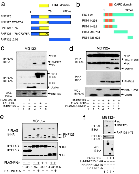
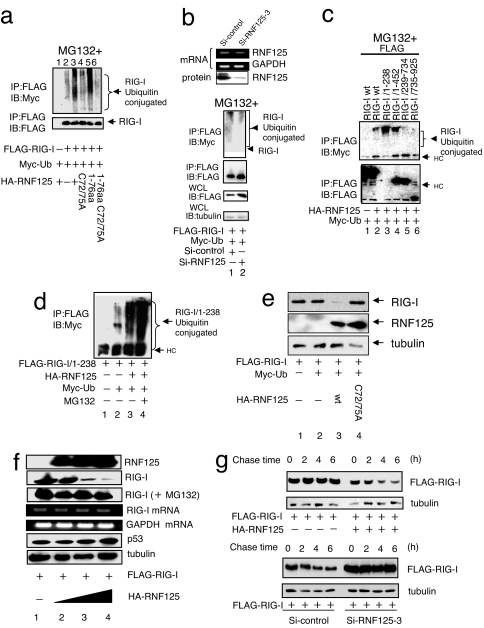
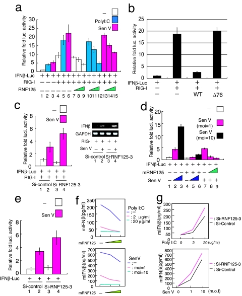
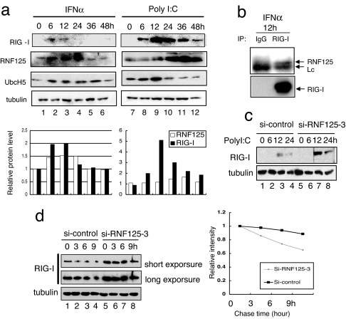
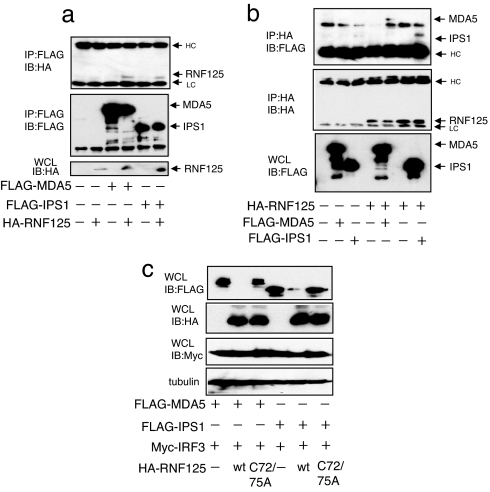
Retinoic acid-inducible gene I (RIG-I) plays a pivotal role in the regulation of cytokine production induced by pathogens. The RIG-I also augments the production of IFN and other cytokines via an amplification circuit. Because the production of cytokines is closely controlled, up- and down-regulation of RIG-I signaling also needs strict regulation. The mechanism of this regulation, however, remains elusive. Here, we found that RIG-I undergoes proteasomal degradation after conjugation to ubiquitin by RNF125. Further, RNF125 conjugates ubiquitin to MDA5, a family protein of RIG-I as well as IPS-1, which is also a downstream protein of RIG-I signaling that results in suppressing the functions of these proteins. Because RNF125 is enhanced by IFN, these functions constitute a negative regulatory loop circuit for IFN production.


Conflict of interest statement

The authors declare no conflict of interest.

Figures

Fig. 1.
Fig. 1.
Association of RNF125 with UbcH8 and RIG-I. (a and b) Schematic structure of RNF125, RIG-I, and their derivatives used in this work. “X” indicates site of cysteine residue substitution with alanine at the 72nd and 75th residues in RNF125. (c and d) Coimmunoprecipitation experiments. The 293FT cells were transfected as indicated. Thirty-six hours after transfection, protein associations were analyzed either by coimmunoprecipitation using anti-FLAG antibody, followed by Western blot using anti-HA antibody (c), or by coimmunoprecipitation using anti-HA antibody, followed by anti-FLAG antibody (d). (e) Analysis of the RIG-I domain that interacts with RNF125. Plasmids expressing various deletion mutants, including amino acids 1–238, 1–452, 239–734, and 735–925 of RIG-I fused to a FLAG epitope tag (b), were transfected into 293FT cells with or without a plasmid encoding HA-RNF125. Complex formation was examined by immunoprecipitation with an anti-FLAG antibody followed by immunoblotting using the anti-HA antibody (Upper). The amount of each RIG-I mutant in the complex is indicated (Lower). (f) Analysis of the association of RIG-I with RNF125 mutants. Plasmids as indicated were transfected into 293FT cells. The lysates were immunoprecipitated by using an anti-FLAG antibody, followed by blotting with the anti-HA antibody (Top). The quantity of FLAG-RIG-I in the immunocomplexes as well as the RNF125 and mutants in whole-cell lysates are also shown in Middle and Bottom, respectively. HC and LC indicate the heavy and light chains of human immunoglobulins, which are also indicated in the following figures. All cells in c–f were treated with MG132.
Fig. 2.
Fig. 2.
Ubiquitin conjugation to RIG-I. (a) Ubiquitin conjugation to RIG-I by WT and mutant RNF125. Plasmids encoding FLAG-RIG-I and Myc-Ub were cotransfected into 292FT cells with a plasmid encoding either WT or mutant RNF125. The RIG-I ubiquitination was monitored by immunoprecipitation. All cells were treated with MG132. (b) Inhibition of RIG-I ubiquitin conjugation after suppression of endogenous RNF125. The siRNF125–3, an siRNA that down-regulates RNF125 mRNA, or a control siRNA were transfected into the 293FT cells. The mRNA and protein levels of RNF125 were monitored by RT-PCR or Western blot, respectively, 36 h after transfection (Upper). The 293FT cells transfected with plasmids encoding FLAG-RIG-I and Myc-Ub were simultaneously treated with control siRNA or siRNF125–3; RIG-I ubiquitination in cell lysates was then analyzed by coimmunoprecipitation. All cells were treated with MG132. The quantity of RIG-I present in the immunoprecipitants was monitored by immunoblot with an anti-FLAG antibody. The total RIG-I present in an aliquot of whole-cell lysate is also shown. Tubulin serves as the control in this analysis (Lower). (c) Analysis of the region for ubiquitin conjugation in RIG-I. Plasmids encoding HA-RNF125 and Myc-Ub were cotransfected into 293FT cells with WT or deletion mutants of FLAG-RIG-I. We analyzed cell lysates harvested 36 h after transfection for interactions between RNF125 and WT or mutant RIG-I by immunoprecipitation (note that the expression level of 1–238 was very low; despite the low levels, however, the interaction with RIG-I was clearly observed). All cells were treated with MG132. (d) Ubiquitin conjugation stimulates RIG-I degradation in a proteasome-dependent manner. Plasmids encoding a FLAG-tagged version of the RIG-I N terminus, FLAG-RIG-I/1–238, HA-RNF125, and Myc-Ub were transfected into 293FT cells as indicated. After culturing with or without MG132, the levels of ubiquitination were analyzed by immunoprecipitation. (e) RNF125 stimulates RIG-I degradation. Plasmids encoding FLAG-RIG-I and Myc-Ub were cotransfected into 293FT cells with WT or a point mutant of HA-RNF125. Cells were harvested 36 h after transfection and were analyzed for proteins by Western blot. (f) RIG-I was degraded by RNF125 in a dose-dependent manner. The 293FT cells were transfected with a plasmid encoding FLAG-RIG-I (0.5 μg) and varying doses of a plasmid encoding HA-RNF125 (0, 0.5, 1, and 2 μg). Half of each cell aliquot was treated with MG132. The levels of both RIG-I mRNA and protein were examined in cells harvested 48 h after transfection. “RIG-I (+MG132)” indicates analysis of the RIG-I protein in cells treated with MG132. As a control, the cellular proteins p53 and tubulin and GAPDH mRNA analyses are shown. (g) RIG-I was degraded by ectopic and endogenous RNF125. A plasmid encoding FLAG-RIG-I with or without a plasmid encoding HA-RNF125 was transfected into 293FT cells. Thirty-six hours after transfection, the cells were treated with CHX (final concentration, 50 μg/ml), and were analyzed by Western blot (Upper). Plasmids encoding FLAG-RIG-I and RNA for si-control or siRNF125–3 were transfected into 293FT cells. Twenty-four hours after transfection, these cells were treated with Poly I:C for 12 h, followed by addition of CHX. Cells were harvested at the indicated times after addition of CHX and analyzed by Western blot (Lower).
Fig. 3.
Fig. 3.
Suppression of RIG-I function by ubiquitination. (a) The RNF125 suppressed the IFNβ-driven luciferase activity, activated by RIG-I (at the left). The 293FT cells were transfected with plasmids encoding RIG-I (50 ng) and IFNβ-luc (50 ng) with varying amounts of a plasmid encoding RNF125 (10, 50, and 100 ng). Twenty-four hours after transfection, cells were treated with polyIC (blue) or infected with Sendai virus (pink). Cells were harvested 12 h after the treatment; luciferase activity in the lysates was then measured. The RNF125 suppressed the production of IFNβ mRNA. (b) The 293FT cells transiently expressing IFNβ-luc, RIG-I, and RNF125WT or Δ76 were analyzed for luciferase activity 36 h after transfection. (c) Effect of siRNF125–3, a small inhibitory RNA specific for RNF125 mRNA, on IFNβ-luc activity and IFNβ level. The 293FT cells were transfected with plasmids encoding IFNβ-luc and RIG-I and treated with control siRNA or siRNF125–3. An aliquot of cells was then infected with Sendai virus. Twenty-four hours after infection, luciferase activity (Left) and IFNβ mRNA levels, assessed by RT-PCR (Right), were measured. (d) Effect of mouse RNF125 on endogenous RIG-I signaling in primary MEFs. The MEFs were transfected with plasmids encoding mouse RNF125 (100 ng in lanes 4, 5, 6, and 8 or 200 ng in lane 9) and IFNβ-luc (50 ng) in combination as indicated. Twelve hours after transfection, cells were infected with Sendai virus at the indicated multiplicity of infection (MOI) or mock infected. Luciferase activity in cell lysates prepared 24 h after treatment was measured. White box, mock infected; pink box, Sendai virus infected with MOI 1; black box, Sendai virus with MOI of 10. (e) The MEFs were transfected with plasmids encoding IFNβ-luc and treated with control siRNA or siRNF125–3 (final concentration of 8 nM). Cells were then infected with Sendai virus at MOI of 1. Twenty-four hours after infection, luciferase activity was measured. (f) The level of IFNβ in culture medium in cells was decreased in an RNF125 dose-dependent manner. Cells transfected with different amounts of plasmids encoding RNF125 were treated with 0 (green), 2 (pink), and 20 (blue) μg/ml of poly I:C (Upper) and with 0 (green), 1 (pink), and 10 MOI (blue) of Sendai virus (Upper). Twelve hours after the treatment, IFNβ in the culture medium was measured by ELISA system. (g) The level of IFNβ was increased in MEFs treated with siRNA for RNF125-specific mRNA. Cells treated with control siRNA (black bars) or siRNA125–3 (pink bars) were treated with different doses of poly I:C or Sendai virus infection and were analyzed for IFNβ in culture medium.
Fig. 4.
Fig. 4.
Induction of RIG-I and the Ubls by IFNα or poly I:C treatment. (a) HepG2 cells were harvested at the indicated times after treatment with IFNα (103 units/ml) or Poly I:C (2 μg/ml) and were analyzed for protein levels by Western blot. As a control, the level of tubulin is shown. The protein level of each band is quantified and graphed. (b) Endogenous association of RIG-I and RNF125. Jurkat cells were incubated with IFNα (1,000 units/ml) and MG132 (final 10 μM). After 12 h, whole-cell lysates were subjected to immunoprecipitation assay using anti-RIG-I antibody or control antibody, followed by immunoblotting for detection of RNF125 and RIG-I. (c) Influence of siRNA specific to RNF125 for RIG-I levels. Cells treated with siRNA were further treated with poly I:C and then harvested at the indicated times after poly I:C treatment. The lysates were analyzed for the levels of endogenous RIG-I. Tubulin acts as a control. (d) HepG2 cells treated with siRNA were further treated with IFNα for 12 h and then harvested at the indicated times after CHX treatment. The lysates were analyzed for the levels of endogenous RIG-I. Tubulin acts as a control. The intensity of each band is quantified and graphed.
Fig. 5.
Fig. 5.
Association of RNF125 with MDA5 and IPS-1. A plasmid encoding FLAG-MDA5 or FLAG-IPS1 was transfected into 293FT cells in the presence or absence of a plasmid encoding HA-RNF125. (a and b) Protein–protein interactions were monitored by coimmunoprecipitation using the anti-FLAG antibody, followed by Western blot with the anti-HA antibody (a) and coimmunoprecipitation using the anti-HA antibody, followed by Western blot with the anti-FLAG antibody (b). All cells were grown in medium containing MG132. (c) MDA5 and IPS1 were also degraded by RNF125. A plasmid encoding Myc-IRF3 and FLAG-MDA5 or FLAG-IPS1 were transfected into 293FT cells with or without a plasmid encoding HA-RNF125 or HA-RNF125 C72/75A. Thirty-six hours after transfection, cells were harvested and were analyzed for protein levels by Western blot.

Similar articles

Cited by

References

    1. Iwasaki A, Medzhitov R. Nat Immunol. 2004;5:987–995. - PubMed
    1. Theofilopoulos AN, Baccala R, Beutler B, Kono DH. Annu Rev Immunol. 2005;23:307–336. - PubMed
    1. Takeda K, Akira S. Int Immunol. 2005;17:1–14. - PubMed
    1. Medzhitov R, Janeway CA., Jr Cell. 1997;91:295–298. - PubMed
    1. Janeway CA, Jr, Medzhitov R. Annu Rev Immunol. 2002;20:197–216. - PubMed

Publication types

MeSH terms