

doi: 10.1523/JNEUROSCI.0325-07.2007.
Conservation of glutamate receptor 2-containing AMPA receptors during long-term potentiation
Affiliations
- PMID: 17460072
- PMCID: PMC6672988
- DOI: 10.1523/JNEUROSCI.0325-07.2007
Item in Clipboard
Conservation of glutamate receptor 2-containing AMPA receptors during long-term potentiation
J Neurosci.
.
Abstract
Long-term potentiation (LTP) at hippocampal synapses is thought to involve the insertion of AMPA receptors into the postsynaptic membrane. Conflicting evidence exists as to whether calcium-permeable receptors are inserted during LTP and whether synaptic activity mediated by the newly inserted AMPA receptors is required to maintain the increase in synaptic strength. Here, we rigorously test these hypotheses and conclude that calcium-permeable AMPA receptors are not inserted during LTP nor does potentiation require ongoing activity to be maintained.
Figures
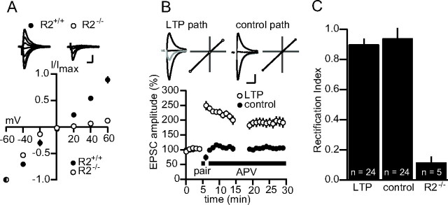
LTP is not associated with a change in the rectification of synaptic AMPAR currents. A, Average I–V plots for synaptic currents in wt (R2+/+; n = 6) and R2−/− (n = 5) pyramidal cells. Top, Example traces. Calibration: 50 pA, 20 ms. B, Summary data in which the I–V profile of synaptic currents was measured in a control and a potentiated pathway (n = 24). Top, Example traces and I–V plots. Gray, Before LTP; black, after LTP. Calibration: 50 pA, 20 ms. C, RIs for a sample of 24 cells between the control and potentiated pathway. The RI for cells in the R2−/− is included for comparison. Error bars represent SEM. In A, mouse brain slices were used; in B and C, rat slices were used. The experiments in B and C were repeated with mouse slices with no apparent difference, and the data were pooled.
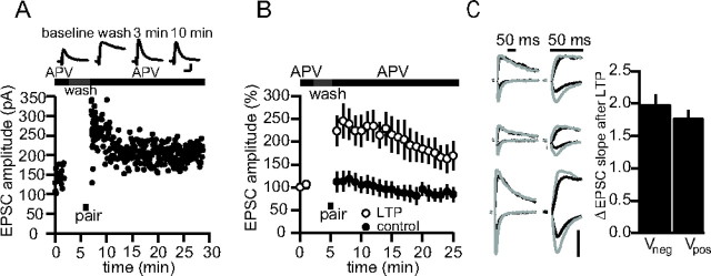
A, Sample experiment in which a cell was voltage clamped at +50 mV during the entire experiment, except during pairing to 0 mV (black bar). Top, Example traces. Calibration: 80 pA, 20 ms. B, Pooled data for nine experiments as in A. C, Sets of sample traces of synaptic responses in three different cells before and after LTP at negative and positive potentials in the absence of APV. Traces are presented on two timescales for comparison. Black, Before LTP; gray, after LTP. Vertical calibration bar: 80 pA. Right, Averaged EPSC slope change for seven such recordings at negative and positive potentials (n = 7; p = 0.43). Error bars represent SEM. Experiments were conducted in rat brain slices.
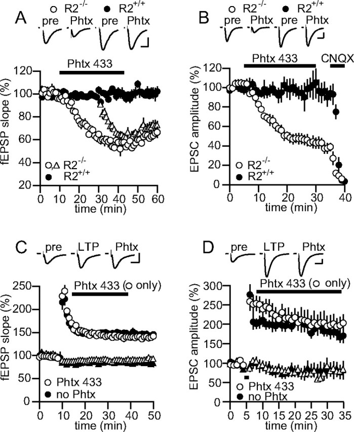
Phtx-433 does not block synaptic currents after LTP. A, Summary data for experiments in which 10 μm Phtx-433 was applied to fEPSPs in R2+/+(n = 4) and R2−/− (n = 4) slices. Open triangles indicate the second pathway in experiments on R2−/− slices (the second pathway for R2+/+ slices has been omitted for clarity). Top, Example traces. Calibration: 0.3 mV, 10 ms. B, Summary data for experiments in which 10 μm Phtx-433 was applied to whole-cell EPSCs in R2+/+(n = 5) and R2−/− (n = 4) slices. Application of 10 μm CNQX at the end of the recording confirmed that the remaining unblocked EPSC was mediated by AMPARs. Top, Example traces. Calibration: 30 pA, 20 ms. C, Summary data for experiments in which 10 μm Phtx-433 (open symbols; n = 10) or no Phtx-433 (closed symbols; n = 8) was applied to fEPSPs after the induction of LTP by tetanization in one pathway (circles) but not the second control (triangles) pathway. Top, Example traces. Calibration: 0.5 mV, 10 ms. D, Summary data for experiments in which 10 μm Phtx-433 (open symbols; n = 9) or no Phtx-433 (closed symbols; n = 8) was applied to whole-cell EPSCs after the induction of LTP by pairing in one pathway (circles) but not the second control pathway (triangles). Top, Example traces. Calibration: 80 pA, 20 ms. All data were collected using mouse brain slices. Error bars represent SEM.
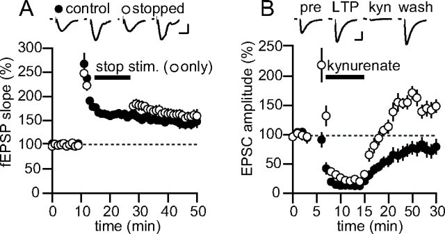
LTP does not require synaptic activity to be maintained. A, Summary data for experiments in which two pathways were tetanized, but one pathway was stopped for 15 min after induction (open symbols). Top, Example traces. n = 6. Calibration: 0.3 mV, 10 ms. B, Summary data for experiments in which kynurenate (1 mm ) was rapidly applied for 7 min after induction of LTP in one pathway (n = 6). Top, Example traces. Calibration: 30 pA, 15 ms. Error bars represent SEM. All data were collected using mouse brain slices.
References
-
- Bellone C, Luscher C. Cocaine triggered AMPA receptor redistribution is reversed in vivo by mGluR-dependent long-term depression. Nat Neurosci. 2006;9:636–641. - PubMed
-
- Clem RL, Barth A. Pathway-specific trafficking of native AMPARs by in vivo experience. Neuron. 2006;49:663–670. - PubMed
-
- Hayashi Y, Shi SH, Esteban JA, Piccini A, Poncer JC, Malinow R. Driving AMPA receptors into synapses by LTP and CaMKII: requirement for GluR1 and PDZ domain interaction. Science. 2000;287:2262–2267. - PubMed
-
- Hollmann M, Heinemann S. Cloned glutamate receptors. Annu Rev Neurosci. 1994;17:31–108. - PubMed
Publication types
MeSH terms
Substances
LinkOut - more resources
Full Text Sources
Other Literature Sources
Molecular Biology Databases