. 2007 Apr 25;27(17):4634-41.
doi: 10.1523/JNEUROSCI.5602-06.2007.

Spinal interneurons that are selectively activated during fictive flexion reflex

Affiliations

Spinal interneurons that are selectively activated during fictive flexion reflex

Ari Berkowitz. J Neurosci. .

Abstract

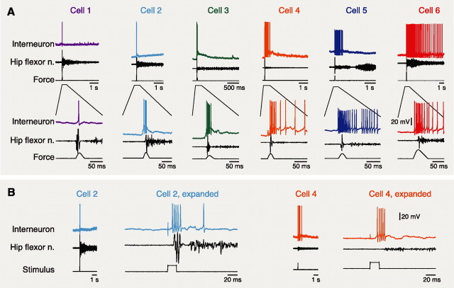
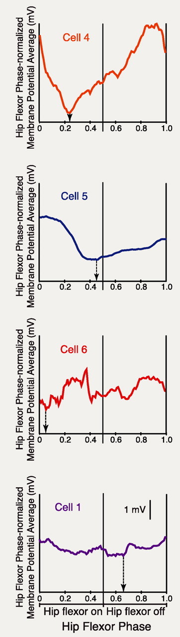
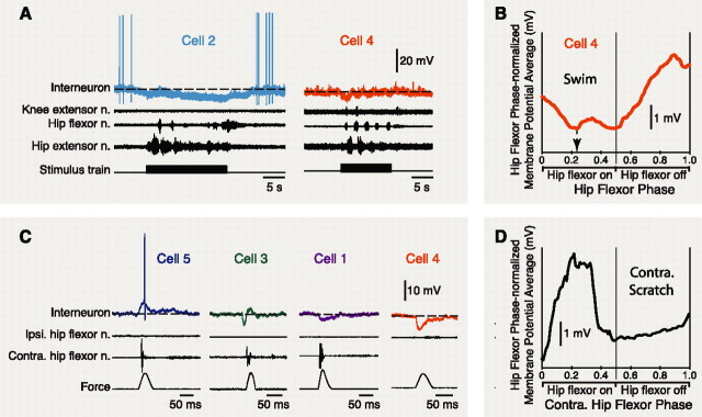
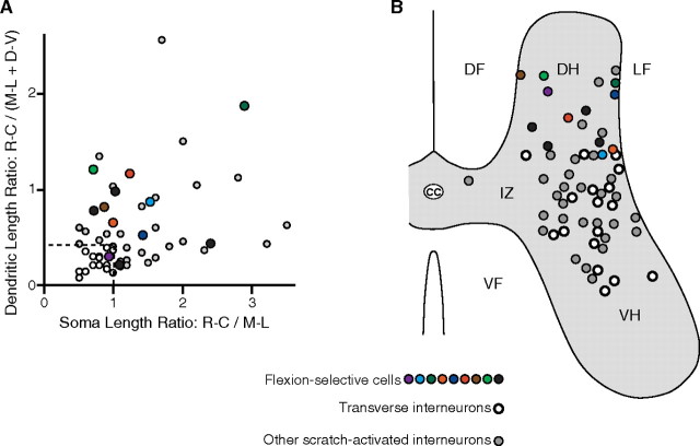
Behavioral choices in invertebrates are mediated by a combination of shared and specialized circuitry, including neurons that are inhibited during competing behaviors. Less is known, however, about the neural mechanisms of behavioral choice in vertebrates. The spinal cord can appropriately select among several types of limb movements, including limb withdrawal (flexion reflex), scratching, and locomotion, and thus is conducive to examination of vertebrate mechanisms of behavioral choice. Flexion reflex can interrupt and reset the rhythm of scratching and locomotion, suggesting that a combination of shared and specialized circuitry contributes to these behaviors, but little is known about the interneurons involved. Here, I used in vivo intracellular recording and dye injection to identify a group of spinal interneurons that are strongly activated during fictive flexion reflex but inhibited during fictive scratching and fictive swimming. These flexion-selective interneurons are typically rhythmically hyperpolarized during fictive scratching and fictive swimming. This hyperpolarization can be maximal during the ipsilateral hip flexor bursts of rhythmic limb motor patterns, although these cells are strongly activated during the ipsilateral hip flexor bursts of fictive flexion reflex. Thus, these interneurons are relatively specialized for fictive limb withdrawal, rather than contributing to the hip flexor phase of multiple types of limb movements. These flexion-selective cells are physiologically and morphologically distinguishable from a recently described group of spinal interneurons (transverse interneurons) that are strongly activated during both fictive flexion reflex and fictive scratching. Thus, spinal interneurons with distinct behavioral roles may to some extent be morphologically distinguishable.


Figures

Figure 1.
Figure 1.
Flexion-selective interneuron responses during ipsilateral fictive flexion reflex. A, Examples of six flexion cells during foot tap-evoked fictive flexion reflex. The bottom sets of traces expand the early portions of the responses. B, Flexion cell responses to electrically evoked fictive flexion reflex for two of the cells shown in A. Note that flexion cell responses can begin within 20 ms of stimulus onset, before the ipsilateral hip flexor nerve burst. In this and subsequent figures, voltage scale bars apply to all cells in that figure panel. n., Nerve.
Figure 2.
Figure 2.
Flexion cell responses during ipsilateral fictive scratching for the cells shown in Figure 1. Bars below each set of traces indicate the period of scratch stimulation. Dashed lines indicate the membrane potential of each cell just before fictive scratching. Note that flexion cells exhibited no response during fictive scratching or were inhibited. n., Nerve.
Figure 3.
Figure 3.
Dual-referent phase-averaged membrane potentials with respect to the ipsilateral hip flexor nerve bursts and interburst intervals during ipsilateral fictive scratching, for four of the cells shown in Figures 1 and 2. Arrows indicate the phase of maximal hyperpolarization in each cell, which could occur during the ipsilateral hip flexor bursts. Cycle periods of all analyzed cycles were 3.17 ± 0.69 s (mean ± SD) for ipsilateral rostral scratching, 2.44 ± 0.99 s for ipsilateral pocket scratching, and 3.54 ± 1.50 s for ipsilateral caudal scratching.
Figure 4.
Figure 4.
Flexion cell responses during additional types of fictive motor patterns. A, Examples of flexion cell responses during fictive forward swimming. B, Dual-referent phase-averaged membrane potential of cell 4 during fictive swimming. Note that its maximal hyperpolarization occurs during the ipsilateral hip flexor bursts. C, Examples of flexion cell responses during contralateral fictive flexion reflex. D, Dual-referent phase-averaged membrane potential with respect to the contralateral hip flexor motor nerve during contralateral fictive scratching for one flexion cell. Cycle periods of all analyzed cycles were 2.26 ± 1.58 s for swimming and 3.12 ± 0.83 s for contralateral scratching. Ipsi., Ipsilateral; Contra., contralateral. n., Nerve.
Figure 5.
Figure 5.
Quantitative analyses of flexion cell membrane potential oscillations during rhythmic motor patterns. A, Peak and trough phases of dual-referent phase-averaged membrane potentials of all flexion cells during ipsilateral fictive scratching. B, Mean ± SEM amplitudes of membrane potential oscillations (peak-to-trough) for all flexion cells studied during ipsilateral fictive scratching (n = 12) and ipsilateral fictive swimming (n = 4), compared with T neurons (n = 16) and non-T neurons (n = 14) during ipsilateral fictive scratching [T neuron and non-T neuron data from Berkowitz et al. (2006)]. Asterisk indicates statistical significance (p = 0.0006).
Figure 6.
Figure 6.
Morphological reconstructions of six flexion cells. A, Cells with relatively simple dendritic branching. B, Cells with relatively complex branching. Note that all cells have rostrocaudally extensive dendrites. DF, Dorsal funiculus; DH, dorsal horn; LF, lateral funiculus; L, left; R, right.
Figure 7.
Figure 7.
Summaries of morphological features of flexion cells. A, Graph of dendritic length ratios versus soma length ratios for all flexion cells (colored and black circles), compared with T neurons (white circles) and other scratch-activated interneurons (gray circles). B, Soma locations for all flexion cells, marked on a schematic cross section and compared with soma locations of T neurons and other scratch-activated interneurons [data on scratch-activated interneurons are from Berkowitz et al. (2006)]. Colors indicate identities of flexion cells shown in previous figures. D-V, Dorsoventral; M-L, mediolateral; R-C, rostrocaudal; cc, central canal; DF, dorsal funiculus; DH, dorsal horn; IZ, intermediate zone; LF, lateral funiculus; VF, ventral funiculus; VH, ventral horn.

Similar articles

Cited by

References

    1. Bannatyne BA, Edgley SA, Hammar I, Jankowska E, Maxwell DJ. Differential projections of excitatory and inhibitory dorsal horn interneurons relaying information from group II muscle afferents in the cat spinal cord. J Neurosci. 2006;26:2871–2880. - PMC - PubMed
    1. Berkowitz A. Broadly tuned spinal neurons for each form of fictive scratching in spinal turtles. J Neurophysiol. 2001;86:1017–1025. - PubMed
    1. Berkowitz A. Both shared and specialized spinal circuitry for scratching and swimming in turtles. J Comp Physiol A Neuroethol Sens Neural Behav Physiol. 2002;188:225–234. - PubMed
    1. Berkowitz A. Physiology and morphology indicate that individual spinal interneurons contribute to diverse limb movements. J Neurophysiol. 2005;94:4455–4470. - PubMed
    1. Berkowitz A. Physiology and morphology of spinal interneurons that are preferentially activated during fictive flexion reflex. Soc Neurosci Abstr. 2006;32:448–26. - PubMed

Publication types

LinkOut - more resources