. 2007 May;48(5):1959-67.
doi: 10.1167/iovs.06-1374.

Bestrophin gene mutations cause canine multifocal retinopathy: a novel animal model for best disease

Affiliations

Bestrophin gene mutations cause canine multifocal retinopathy: a novel animal model for best disease

Karina E Guziewicz et al. Invest Ophthalmol Vis Sci. 2007 May.

Abstract

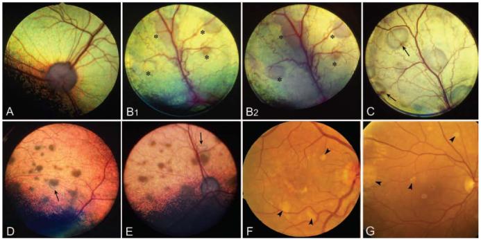
Purpose: Canine multifocal retinopathy (cmr) is an autosomal recessive disorder of multiple dog breeds. The disease shares a number of clinical and pathologic similarities with Best macular dystrophy (BMD), and cmr is proposed as a new large animal model for Best disease.

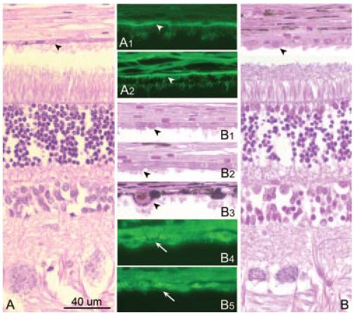
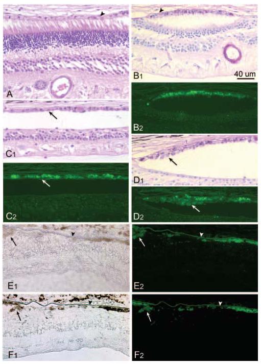
Methods: cmr was characterized by ophthalmoscopy and histopathology and compared with BMD-affected patients. BEST1 (alias VMD2), the bestrophin gene causally associated with BMD, was evaluated in the dog. Canine ortholog cDNA sequence was cloned and verified using RPE/choroid 5'- and 3'-RACE. Expression of the canine gene transcripts and protein was analyzed by Northern and Western blotting and immunocytochemistry. All exons and the flanking splice junctions were screened by direct sequencing.

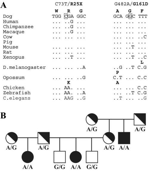
Results: The clinical phenotype and pathology of cmr closely resemble lesions of BMD. Canine VMD2 spans 13.7 kb of genomic DNA on CFA18 and shows a high level of conservation among eukaryotes. The transcript is predominantly expressed in RPE/choroid and encodes bestrophin, a 580-amino acid protein of 66 kDa. Immunocytochemistry of normal canine retina demonstrated specific localization of protein to the RPE basolateral plasma membranes. Two disease-specific sequence alterations were identified in the canine VMD2 gene: a C(73)T stop mutation in cmr1 and a G(482)A missense mutation in cmr2.

Conclusions: The authors propose these two spontaneous mutations in the canine VMD2 gene, which cause cmr, as the first naturally occurring animal model of BMD. Further development of the cmr models will permit elucidation of the complex molecular mechanism of these retinopathies and the development of potential therapies.


Figures

Figure 1
Figure 1
Fundus photographs illustrating the salient retinal findings of cmr1 and cmr2 and multifocal Best disease. (A) Healthy control showing that the fundus is divided into the superior tapetal and the inferior nontapetal regions. In the tapetal region, the retinal pigment epithelium in nonpigmented, and overlies the brightly reflective tapetal layer of the choroid. (B) Great Pyrenees with cmr1 at 5 (B1) and 8 (B2) months of age. Retinal lesions (*) are more distinct at 8 months of age. (C) Eighteen-month-old Coton de Tulear with cmr2. The fluid in the subretinal elevations is clear, though two lesions (arrows) show accumulation of a tan-pink material in the dependent portion of the blebs. (D–E) Fifteen-month-old Coton de Tulear with cmr2. Multifocal retinal lesions have tan-pink brown subretinal material. Adjacent to several lesions (arrows) the subretinal fluid is serous. (F–G) Multifocal Best disease in humans shown in 30° and 60° fundus photographs of patients with heterozygous A243T (F, 30°) and Y227N (G, 60°) mutations. Multifocal lesions (arrowheads) are present in both eyes.
Figure 2
Figure 2
Samples of normal (A, 1 year) and affected (VMD2 stop mutation; B, 4 years) Great Pyrenees dogs. The normal RPE is not hypertrophied (A) and shows uniform autofluorescence in the basal zone (arrowheads,A1,A2). The affected RPE is hypertrophied (arrowheads; B, B1B3), and bright autofluorescent granules occupy the cytoplasm (white arrows; B4, B5).
Figure 3
Figure 3
Retinal pathology in cmr1 and Best disease. (AD) Photomicrographs from archival sample of 10-year-old affected (VMD2stop mutation) Great Pyrenees dog. The retina in localized areas of the posterior pole is normal (A), though most areas show outer retinal atrophy with RPE cells hypertrophy (BD). The RPE cells have granular to tan-brown cytoplasmic inclusions (B1, C1, D1) that are PAS+ (B1, D1) and autofluorescent (B2, C2, D2). Proliferation of RPE leads to the formation of cell nests (D1, D2). (EF) Photomicrographs of the juxtafoveal region of the retina from an 86-year-old patient with a T6R mutation in VMD2. Clusters of pigmented cells are present (arrows), and extensive intracytoplasmic lipofuscin accumulation results in bright granular autofluorescence. Areas of photoreceptor loss are associated with regions of RPE dropout, but BM is intact (arrowheads).
Figure 4
Figure 4
Sequence divergence of VMD2 in 14 different species. The neighbor-joining tree was derived from a bootstrapped (n = 1000) F84 model using experimental data (black) and predicted gene sequences, where applicable (gray).
Figure 5
Figure 5
Expression of VMD2-specific transcripts. (A) VMD2 expression in eight canine tissues by RT-PCR. PCR products in RPE/choroid, retina and brain contained the cDNA fragment of expected size (329 bp); GAPDH control. (B) Northern blot of VMD2 expression detected the approximately 2.1-kb transcript only in RPE/choroid. (C) Western blot analysis with monoclonal E6–6 antibody showed a single band of approximately 60 kDa in the normal canine RPE/choroid sample.
Figure 6
Figure 6
Immunohistochemistry showing the localization of bestrophin (red) in the RPE of C. familiaris (A1A2), M. fascicularis (B), and F. catus (C). The protein is highly expressed in the basolateral membranes of the retinal pigment epithelium (RPE) but not in other retinal layers or the choroid. Nuclei are stained blue with DAPI. Dashed line (B): the apposition of the artifactually separated retina to the RPE. ONL, outer nuclear layer; INL, inner nuclear layer. Scale bars: 80 μm (A1, B, C); 20 μm (A2).
Figure 7
Figure 7
Identification and cosegregation of the canine VMD2 gene mutations. (A) Conservation of the sequences surrounding the mutation C73T (R25Stop) of cmr1 and the G482A (G161D) change of cmr2 in 14 VMD2 orthologs. (B) Part of the Coton de Tulear pedigree segregating cmr2 with recessive inheritance of mutant allele (G, normal allele; A, mutant allele). Clinically evident fundus abnormalities are noticeable in homozygous mutant (A/A) dogs but not in heterozygotes (A/G).

References

    1. Akhmedov NB, Piriev NI, Chang B, et al. A deletion in a photoreceptor-specific nuclear receptor mRNA causes retinal degeneration in the rd7 mouse. Proc Natl Acad Sci USA. 2000;97:5551–5556. - PMC - PubMed
    1. D'Cruz PM, Yasumura D, Weir J, et al. Mutation of the receptor tyrosine kinase gene Mertk in the retinal dystrophic RCS rat. Hum Mol Genet. 2000;9:645–651. - PubMed
    1. Kijas JW, Cideciyan AV, Aleman TS, et al. Naturally occurring rhodopsin mutation in the dog causes retinal dysfunction and degeneration mimicking human dominant retinitis pigmentosa. Proc Natl Acad Sci USA. 2002;99:6328–6333. - PMC - PubMed
    1. Cideciyan AV, Jacobson SG, Aleman TS, et al. In vivo dynamics of retinal injury and repair in the rhodopsin mutant dog model of human retinitis pigmentosa. Proc Natl Acad Sci USA. 2005;102:5233–5238. - PMC - PubMed
    1. Aguirre GD, Acland GM. Models, mutants and man: searching for unique phenotypes and genes in the dog model of inherited retinal degeneration. In: Ostrander EA, Giger U, Lindblad-Toh K, editors. The Dog and Its Genome. Cold Spring Harbor Laboratory Press; Cold Spring Harbor, NY: 2006.

Publication types

MeSH terms