

Comparative analysis of human conjunctival and corneal epithelial gene expression with oligonucleotide microarrays
- PMID: 17460260
- PMCID: PMC2909883
- DOI: 10.1167/iovs.06-0998
Comparative analysis of human conjunctival and corneal epithelial gene expression with oligonucleotide microarrays
Abstract
Purpose: To determine global mRNA expression levels in corneal and conjunctival epithelia and identify transcripts that exhibit preferential tissue expression.
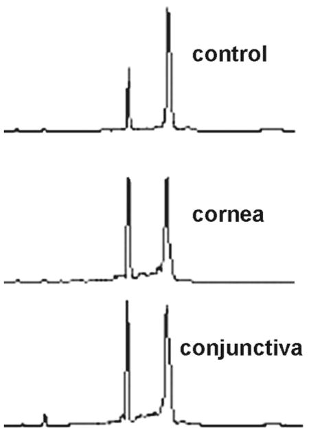
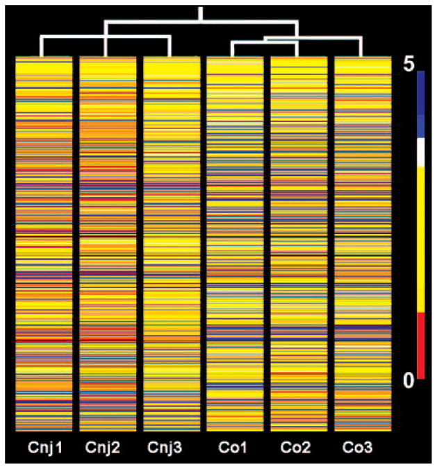
Methods: cDNA samples derived from human conjunctival and corneal epithelia were hybridized in three independent experiments to a commercial oligonucleotide array representing more than 22,000 transcripts. The resultant signal intensities and microarray software transcript present/absent calls were used in conjunction with the local pooled error (LPE) statistical method to identify transcripts that are preferentially or exclusively expressed in one of the two tissues at significant levels (expression >1% of the beta-actin level). EASE (Expression Analysis Systematic Explorer software) was used to identify biological systems comparatively overrepresented in either epithelium. Immuno-, and cytohistochemistry was performed to validate or expand on selected results of interest.
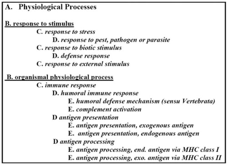
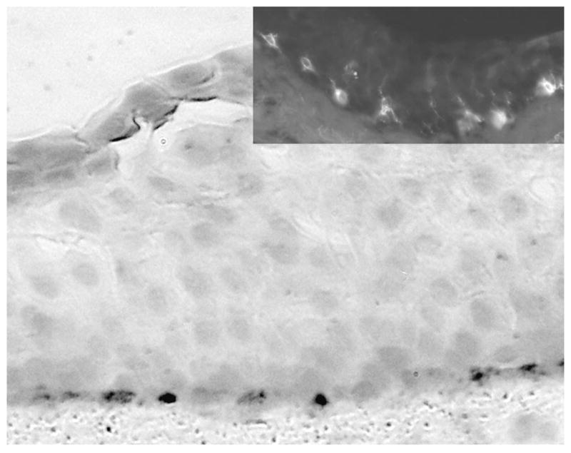
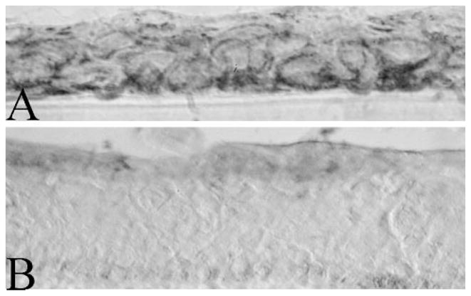
Results: The analysis identified 332 preferential and 93 exclusive significant corneal epithelial transcripts. The corresponding numbers of conjunctival epithelium transcripts were 592 and 211, respectively. The overrepresented biological processes in the cornea were related to cell adhesion and oxiredox equilibria and cytoprotection activities. In the conjunctiva, the biological processes that were most prominent were related to innate immunity and melanogenesis. Immunohistochemistry for antigen-presenting cells and melanocytes was consistent with these gene signatures. The transcript comparison identified a substantial number of genes that have either not been identified previously or are not known to be highly expressed in these two epithelia, including testican-1, ECM1, formin, CRTAC1, and NQO1 in the cornea and, in the conjunctiva, sPLA(2)-IIA, lipocalin 2, IGFBP3, multiple MCH class II proteins, and the Na-Pi cotransporter type IIb.
Conclusions: Comparative gene expression profiling leads to the identification of many biological processes and previously unknown genes that are potentially active in the function of corneal and conjunctival epithelia.
Figures

Similar articles
-
Keratin 13 is a more specific marker of conjunctival epithelium than keratin 19.Mol Vis. 2011;17:1652-61. Epub 2011 Jun 18. Mol Vis. 2011. PMID: 21738394 Free PMC article.
-
Molecular profiling of conjunctival epithelial side-population stem cells: atypical cell surface markers and sources of a slow-cycling phenotype.Invest Ophthalmol Vis Sci. 2009 Sep;50(9):4162-72. doi: 10.1167/iovs.08-2861. Epub 2009 Mar 25. Invest Ophthalmol Vis Sci. 2009. PMID: 19324848 Free PMC article.
-
MUC16 mucin is expressed by the human ocular surface epithelia and carries the H185 carbohydrate epitope.Invest Ophthalmol Vis Sci. 2003 Jun;44(6):2487-95. doi: 10.1167/iovs.02-0862. Invest Ophthalmol Vis Sci. 2003. PMID: 12766047
-
Clusters of corneal epithelial cells reside ectopically in human conjunctival epithelium.Invest Ophthalmol Vis Sci. 2006 Apr;47(4):1359-67. doi: 10.1167/iovs.05-1084. Invest Ophthalmol Vis Sci. 2006. PMID: 16565369
-
Role of mucins in the function of the corneal and conjunctival epithelia.Int Rev Cytol. 2003;231:1-49. doi: 10.1016/s0074-7696(03)31001-0. Int Rev Cytol. 2003. PMID: 14713002 Review.
Cited by
-
Ocular surface development and gene expression.J Ophthalmol. 2013;2013:103947. doi: 10.1155/2013/103947. Epub 2013 Feb 21. J Ophthalmol. 2013. PMID: 23533700 Free PMC article.
-
Use of bioanalyzer electropherograms for quality control and target evaluation in microarray expression profiling studies of ocular tissues.J Ocul Biol Dis Infor. 2009 Dec 12;2(4):243-249. doi: 10.1007/s12177-009-9046-2. J Ocul Biol Dis Infor. 2009. PMID: 20157354 Free PMC article.
-
Keratin 13 is a more specific marker of conjunctival epithelium than keratin 19.Mol Vis. 2011;17:1652-61. Epub 2011 Jun 18. Mol Vis. 2011. PMID: 21738394 Free PMC article.
-
Comparative transcriptional profiling of the limbal epithelial crypt demonstrates its putative stem cell niche characteristics.BMC Genomics. 2010 Sep 29;11:526. doi: 10.1186/1471-2164-11-526. BMC Genomics. 2010. PMID: 20920242 Free PMC article.
-
Regeneration of the corneal epithelium with conjunctival epithelial equivalents generated in serum- and feeder-cell-free media.Mol Vis. 2013 Dec 16;19:2542-50. eCollection 2013. Mol Vis. 2013. PMID: 24357922 Free PMC article.
References
-
- Arcellana-Panlilio M, Robbins SM. Cutting-edge technology. I. Global gene expression profiling using DNA microarrays. Am J Physiol. 2002;282:G397–G402. - PubMed
-
- Verducci JS, Melfi VF, Lin S, Wang Z, Roy S, Sen CK. Microarray analysis of gene expression: considerations in data mining and statistical treatment. Physiol Genomics. 2006;25:355–363. - PubMed
-
- Schena M, Shalon D, Davis RW, Brown PO. Quantitative monitoring of gene expression patterns with a complementary DNA microarray. Science. 1995;270:467–470. - PubMed
-
- Sun CC, Su Pang JH, Cheng CY, et al. Interleukin-1 receptor antagonist (IL-1RA) prevents apoptosis in ex vivo expansion of human limbal epithelial cells cultivated on human amniotic membrane. Stem Cells. 2006;24:2130–2139. - PubMed
Publication types
MeSH terms
Substances
Grants and funding
LinkOut - more resources
Full Text Sources
Molecular Biology Databases
Research Materials
Miscellaneous
