

Minimally activated CD8 autoreactive T cells specific for IRBP express a high level of Foxp3 and are functionally suppressive
- PMID: 17460277
- PMCID: PMC2577316
- DOI: 10.1167/iovs.06-1189
Minimally activated CD8 autoreactive T cells specific for IRBP express a high level of Foxp3 and are functionally suppressive
Abstract
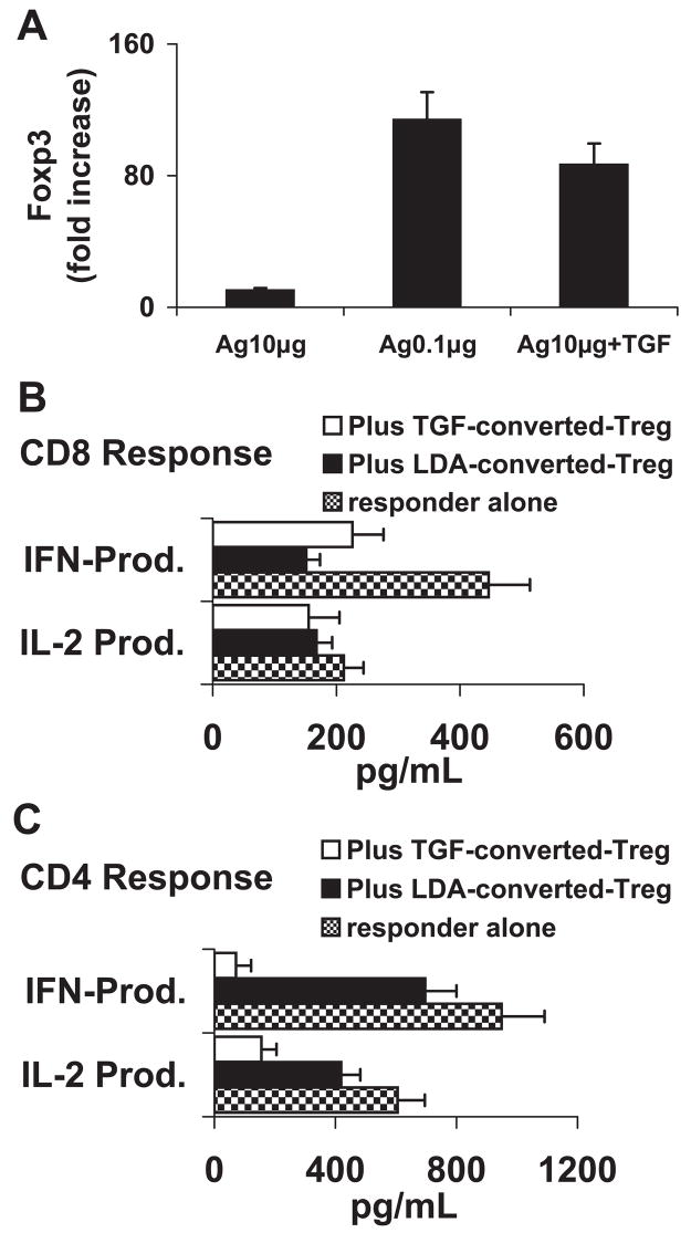
Purpose: Results in previous reports have demonstrated that immunization of the EAU-prone B6 mouse activates both CD4 and CD8 IRBP-specific T cells. The purpose of this study was to investigate structural and functional differences between CD4 and CD8 autoreactive T cells activated by the uveitogenic peptide.
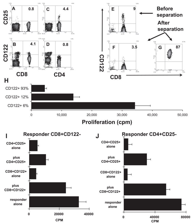
Methods: Purified CD4 and CD8 isolated from B6 mice immunized with an uveitogenic peptide, interphotoreceptor retinoid-binding protein (IRBP)1-20, were stimulated in vitro with various doses of immunizing peptide. The activated T cells were determined for cytokine production, expression of Foxp3, and suppressor activity.
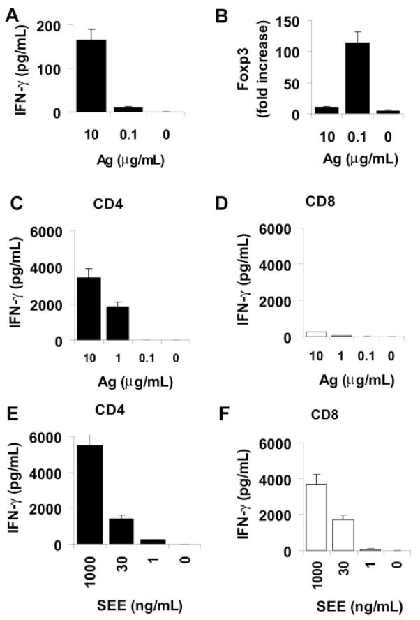
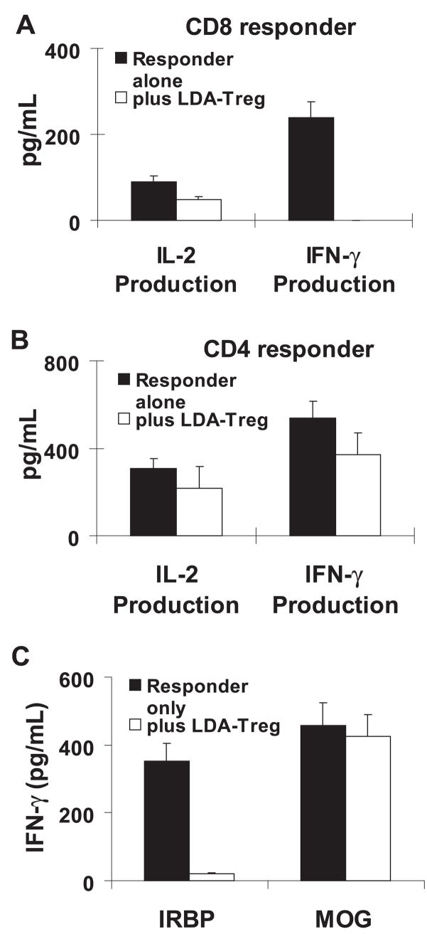
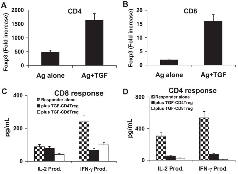
Results: CD4 autoreactive T cells underwent full activation when stimulated with high or medium concentrations of immunizing peptide, whereas a high dose of antigenic peptide resulted in only modest activation of CD8 autoreactive T cells. When stimulated by a low dose (<0.1 microg/mL) of antigen or by of a high dose of antigen and a small amount of TGF-beta1, the minimally activated CD8 T cells expressed a high level of Foxp3 and gained suppressor function.
Conclusions: Minimally activated CD8 autoreactive T cells can be functionally suppressive and may neutralize the tissue-damaging effect of the CD4 autoreactive T cells.
Figures
Similar articles
-
Characterization of IL-17+ interphotoreceptor retinoid-binding protein-specific T cells in experimental autoimmune uveitis.Invest Ophthalmol Vis Sci. 2007 Sep;48(9):4153-61. doi: 10.1167/iovs.07-0251. Invest Ophthalmol Vis Sci. 2007. PMID: 17724201 Free PMC article.
-
In vitro activation of CD8 interphotoreceptor retinoid-binding protein-specific T cells requires not only antigenic stimulation but also exogenous growth factors.J Immunol. 2006 Apr 15;176(8):5006-14. doi: 10.4049/jimmunol.176.8.5006. J Immunol. 2006. PMID: 16585597
-
A shared epitope of the interphotoreceptor retinoid-binding protein recognized by the CD4+ and CD8+ autoreactive T cells.J Immunol. 2005 Aug 1;175(3):1851-7. doi: 10.4049/jimmunol.175.3.1851. J Immunol. 2005. PMID: 16034128 Free PMC article.
-
Suppression of established experimental autoimmune uveitis by anti-LFA-1alpha Ab.Invest Ophthalmol Vis Sci. 2007 Jun;48(6):2667-75. doi: 10.1167/iovs.06-1383. Invest Ophthalmol Vis Sci. 2007. PMID: 17525198 Free PMC article.
-
Repertoire analysis and new pathogenic epitopes of IRBP in C57BL/6 (H-2b) and B10.RIII (H-2r) mice.Invest Ophthalmol Vis Sci. 2008 May;49(5):1946-56. doi: 10.1167/iovs.07-0868. Invest Ophthalmol Vis Sci. 2008. PMID: 18436827 Free PMC article.
Cited by
-
Activated gammadelta T cells promote the activation of uveitogenic T cells and exacerbate EAU development.Invest Ophthalmol Vis Sci. 2011 Jul 29;52(8):5920-7. doi: 10.1167/iovs.10-6758. Invest Ophthalmol Vis Sci. 2011. PMID: 21296823 Free PMC article.
-
Characterization of a New Epitope of IRBP That Induces Moderate to Severe Uveoretinitis in Mice With H-2b Haplotype.Invest Ophthalmol Vis Sci. 2015 Aug;56(9):5439-49. doi: 10.1167/iovs.15-17280. Invest Ophthalmol Vis Sci. 2015. PMID: 26284549 Free PMC article.
-
Upregulation of CD94 on CD8+T cells in anterior chamber-associated immune deviation.BMC Immunol. 2008 Sep 25;9:53. doi: 10.1186/1471-2172-9-53. BMC Immunol. 2008. PMID: 18816417 Free PMC article.
-
TNF inhibitors target a mevalonate metabolite/TRPM2/calcium signaling axis in neutrophils to dampen vasculitis in Behçet's disease.Nat Commun. 2024 Oct 26;15(1):9261. doi: 10.1038/s41467-024-53528-3. Nat Commun. 2024. PMID: 39461948 Free PMC article.
-
Pivotal roles of T-helper 17-related cytokines, IL-17, IL-22, and IL-23, in inflammatory diseases.Clin Dev Immunol. 2013;2013:968549. doi: 10.1155/2013/968549. Epub 2013 Jul 14. Clin Dev Immunol. 2013. PMID: 23956763 Free PMC article. Review.
References
-
- Katz JD, Wang B, Haskins K, et al. Following a diabetogenic T cell from genesis through pathogenesis. Cell. 1993;74:1089–1100. - PubMed
-
- Goverman J, Woods A, Larson L, et al. Transgenic mice that express a myelin basic protein-specific T cell receptor develop spontaneous autoimmunity. Cell. 1993;72:551–560. - PubMed
-
- Shevach EM. Regulatory T cells in autoimmunity. Annu Rev Im-munol. 2000;18:423–449. - PubMed
-
- Suripayer E, Amar AZ, Thornton AM, Shevach EM. CD4+CD25+ T cells inhibit both the induction and effector function of autoreac-tive T cells and represent a unique lineage of immunoregulatory cells. J Immunol. 1998;160:1212–1218. - PubMed
-
- Sun D, Qin Y, Chluba J, et al. Suppression of experimentally-induced autoimmune encephalomyelitis by cytolytic T-T cell interactions. Nature. 1988;332:843–846. - PubMed
Publication types
MeSH terms
Substances
Grants and funding
LinkOut - more resources
Full Text Sources
Research Materials
