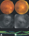
Laser photocoagulation combined with intravitreal triamcinolone acetonide injection in proliferative diabetic retinopathy with macular edema
- PMID: 17460426
- PMCID: PMC2629684
- DOI: 10.3341/kjo.2007.21.1.11
Laser photocoagulation combined with intravitreal triamcinolone acetonide injection in proliferative diabetic retinopathy with macular edema
Abstract
Purpose: To evaluate therapeutic effects and usefulness of a combination treatment of intravitreal injection of triamcinolone acetonide (IVTA) and panretinal photocoagulation (PRP) in patients with clinically significant macular edema secondary to proliferative diabetic retinopathy (PDR).
Methods: Visual acuity test, fundoscopy, fluorescein angiography, and optical coherence tomography (OCT) were taken in 20 patients (20 eyes) of macular edema and PDR. A combination of intravitreal injection of triamcinolone acetonide and PRP was performed in 10 patients (10 eyes) and a combination of focal or grid laser photocoaqulation and PRP in the remaining 10 eyes. The postoperative outcomes were compared between the two combination treatments by best corrected visual acuity (BCVA), tonometry, fluorescein angiography, and OCT at 2 weeks, 1, 2, and 3 months.
Results: Average BCVA (log MAR) significantly improved from preoperative 0.56-/+0.20 to 0.43-/+0.08 at 1 month (P=0.042) and it was maintained until 3 months after a combination of IVTA and PRP in 10 eyes (P=0.007). The thickness of fovea decreased from average 433.3-/+114.9 micrometer to average 279.5-/+34.1 micrometer at 2 weeks after combined treatment of IVTA and PRP (P=0.005), which was significantly maintained until 3 months, but there was a transient visual disturbance and no significant difference in thickness of the fovea before and after treatment in the groups with PRP and focal or grid laser photocoagulation.
Conclusions: A combination of IVTA and PRP might be an effective treatment modality in the treatment of macular edema and PDR and prevent the subsequent PRP-induced macular edema result in visual dysfunction. In combination with PRP, IVTA might be more effective than focal or grid laser photocoagulation and PRP for reducing diabetic macular edema and preventing aggravation of macular edema without transient visual disturbance in patients requiring immediate PRP.
Figures


References
-
- Aiello LM. Perspectives on diabetic retinopathy. Am J Ophthalmol. 2003;136:122–135. - PubMed
-
- Akduman L, Olk RJ. Laser photocoagulation of diabetic macular edema. Ophthalmic Surg Lasers. 1997;28:387–408. - PubMed
-
- Early Treatment Diabetic Retinopathy Study Research Group. Photocoagulation for diabetic macular edema. Early Treatment Diabetic Retinopathy Study report number 1. Arch Ophthalmol. 1985;103:1796–1806. - PubMed
-
- Early Treatment Diabetic Retinopathy Study Research Group. Treatment techniques and clinical guidelines for photocoagulation of diabetic macular edema. Early Treatment Diabetic Retinopathy Study report number 2. Ophthalmology. 1987;94:761–774. - PubMed
-
- Ferris FL, Podgor MJ, Davis MD. Macular edema in diabetic retinopathy study patients Diabetic Retinopathy Study Group report number 12. Ophthalmology. 1987;94:754–760. - PubMed
MeSH terms
Substances
LinkOut - more resources
Full Text Sources
Medical
Research Materials

