

Endogenous aeromonas hydrophila endophthalmitis in an immunocompromised patient
- PMID: 17460432
- PMCID: PMC2629690
- DOI: 10.3341/kjo.2007.21.1.45
Endogenous aeromonas hydrophila endophthalmitis in an immunocompromised patient
Abstract
Purpose: To report a case of endogenous endophthalmitis due to Aeromonas hydrophila in a patient with distal common bile duct carcinoma and biliary sepsis.
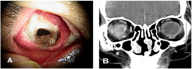
Methods: A 72-year-old woman with distal common bile duct carcinoma, obstructive jaundice, diabetes mellitus, and hypertension had a 1-day history of blurred vision, redness, and eye discharges in the right eye. An ophthalmic examination showed no light perception vision, increased intraocular pressure, severe corneal edema, severe anterior chamber reaction, exudative membranes on the anterior lens surface, and severe vitreal reaction. There was no ocular history of trauma, infection, or surgery in either eye.
Results: Under the impression of endogenous bacterial endophthalmitis, immediate intraocular cultures and intravitreal antibiotic injections were performed, but the anterior chamber reaction, and the ultrasonogram findings were deteriorated. Evisceration was undertaken because of extrusion of the intraocular contents, and Aeromonas hydrophila was isolated by intraocular culture.
Conclusions: Endogenous endophthalmitis due to Aeromonas hydrophila is rare, but has a rapid clinical course and a poor prognosis, despite of prompt diagnosis and management.
Figures


References
-
- Frieling JS, Rosenberg R, Edelstein M, et al. Endogenous Aeromonas hydrophila endophthalmitis. Ann Ophthalmol. 1989;21:117–118. - PubMed
-
- Tamura T, Hida T. A case of endogenous Aeromonas hydrophila endophthalmitis. Nippon Ganka Gakkai Zasshi. 2003;107:535–537. - PubMed
-
- Jackson TL, Eykyn SJ, Graham EM, Stanford MS. Endogenous bacterial endophthalmitis: a 17-year prospective series and review of 267 reported cases. Surv Ophthalmol. 2003;48:403–423. - PubMed
-
- Cohen KL, Holyk PR, McCarthy LR, Peiffer RL. Aeromonas hydrophila and Plesiomonas shigelloides endophthalmitis. Am J Ophthalmol. 1983;96:403–404. - PubMed
-
- Feaster FT, Nisbet R, Barber JC. Aeromonas hydrophila corneal ulcer. Am J Ophthalmol. 1978;85:114–117. - PubMed
Publication types
MeSH terms
Substances
LinkOut - more resources
Full Text Sources
