. 2007 Apr 25;2(4):e388.
doi: 10.1371/journal.pone.0000388.

Enriched monolayer precursor cell cultures from micro-dissected adult mouse dentate gyrus yield functional granule cell-like neurons

Affiliations

Enriched monolayer precursor cell cultures from micro-dissected adult mouse dentate gyrus yield functional granule cell-like neurons

Harish Babu et al. PLoS One. .

Abstract

Background: Stem cell cultures are key tools of basic and applied research in Regenerative Medicine. In the adult mammalian brain, lifelong neurogenesis originating from local precursor cells occurs in the neurogenic regions of the hippocampal dentate gyrus. Despite widespread interest in adult hippocampal neurogenesis and the use of mouse models to study it, no protocol existed for adult murine long-term precursor cell cultures with hippocampus-specific differentiation potential.

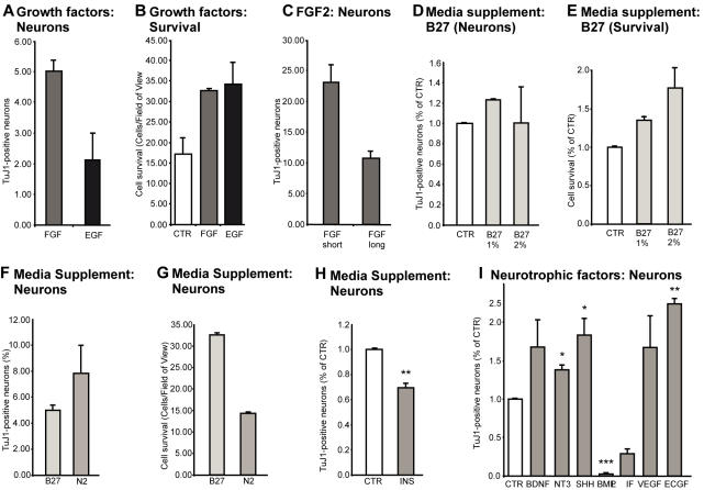
Methodology/principal findings: We describe a new strategy to obtain serum-free monolayer cultures of neural precursor cells from microdissected dentate gyrus of adult mice. Neurons generated from these adherent hippocampal precursor cell cultures expressed the characteristic markers like transcription factor Prox1 and showed the TTX-sensitive sodium currents of mature granule cells in vivo. Similar to granule cells in vivo, treatment with kainic acid or brain derived neurotrophic factor (BDNF) elicited the expression of GABAergic markers, further supporting the correspondence between the in vitro and in vivo phenotype. When plated as single cells (in individual wells) or at lowest density for two to three consecutive generations, a subset of the cells showed self-renewal and gave rise to cells with properties of neurons, astrocytes and oligodendrocytes. The precursor cell fate was sensitive to culture conditions with their phenotype highly influenced by factors within the media (sonic hedgehog, BMP, LIF) and externally applied growth factors (EGF, FGF2, BDNF, and NT3).

Conclusions/significance: We report the conditions required to generate adult murine dentate gyrus precursor cell cultures and to analyze functional properties of precursor cells and their differentiated granule cell-like progeny in vitro.


Conflict of interest statement

Competing Interests: The authors have declared that no competing interests exist.

Figures

Figure 1
Figure 1
Isolation of endogenous precursor cells from the adult mouse dentate gyrus. A; Coronal 300 µm vibratome section through the brain of a Nestin-GFP transgenic mouse. B; As a second step, the dentate gyrus was isolated from the rest of the tissue by microdissection. The dissected dentate gyrus is shown at the top. C; The dissected dentate gyrus, as depicted in B, was further trimmed by placing two cuts that separate the dentate gyrus from ventricular tissue (of the 3rd ventricle) and area CA3 (see also D). Nestin-GFP expression in the subgranular zone is seen with high concentration at the medial tip of the granule cell layer. D; Schematic drawing depicting the dissected region that was used for isolation of the precursor cells. The localization of the potentially contaminating ventricular areas is highlighted in red. The dashed lines indicate the cuts described in C. E; The tissue was homogenized and separated on a Percoll density gradient. Proliferating precursors were present in the region with density>1.053 gm/ml (>22% Percoll). The Erythrocytes were enriched in the region with density>1.075 gm/ml (>65% Percoll). F; phase contrast image of mouse precursor cells grown as adherent cultures. Note the phase bright appearance that is characteristic of proliferating precursor cells. G; Incubation with BrdU (20 µM) revealed that isolated nestin-expressing cells were proliferating. Nestin (Green), BrdU (Red).
Figure 2
Figure 2
Proliferating cells in culture showed features of radial glia and upon differentiation generated neurons and astroglial cells. A–D; Proliferating cultures expressed Nestin (A), RC2 (B), BLBP (C) D; Overlay of the three markers Nestin (green), RC2 (red) and BLBP (blue). E–G; Precursor cells also immunostained for GLAST (E) and Sox2 (F), overlay of GLAST and Sox2 (G). H; RT-PCR analysis of mRNA from precursor cells further corroborated the suggestive radial glia-like phenotype: Nestin, GFAP, Vimentin, BLBP, Pax6, Sox2. I,J; Under differentiation conditions precursor cells generated neurons and astroglia (bIII-tubulin (green) GFAP (blue) and NG2 (oligodendrocyte precursors). K,L; Addition of BrdU before the transfer to differentiation conditions revealed BrdU (green) in neurons expressing Map2ab (red), confirming that the neuron-like cells originated from a proliferating precursor cell.
Figure 3
Figure 3
Differentiated neurons originating from hippocampal precursor cells showed features of dentate gyrus granule cells. After differentiation the cells were fixed and stained for markers characteristic of dentate gyrus granule cells. A; β-III-tubulin (green), Prox1(red). B; β-III-tubulin (green), Calbindin (Red). C; β-III-tubulin (Green), Synaptoporin (Red). D; β-III-tubulin (Green), NeuN (Red). E; Differentiation led to up-regulation of molecular neuronal markers and down-regultion of precursor cell markers. Proliferating cultures (day 0) were allowed to differentiate and mRNA was extracted after different times. Differentiation led to up-regulation of NeuroD1, mash1, VGLUT1 transcripts, whereas Nestin was down-regulated. F; Up-regulation of GAD67 by differentiated neurons. Neurons differentiated for 10–12 days were washed once with medium and subjected to a 2.5 h stimulation of kainate (KA, 10 µM) or rhBDNF (100 ng/ml) in fresh medium and subsequently fixed and evaluated. GAD67 (red) was upregulated in β-III-tubulin-positive cells (green), a property associated with granule neurons in vivo. The images were captured with identical laser settings at the confocal microscope. G; Electrical properties of cultured dentate gyrus precursor cells and differentiated neurons. Membrane properties of cultured dentate gyrus precursor cells and neurons derived from them were measured while voltage-clamped to −60mV. G, H, J, K; Typical membrane currents recorded by series of depolarizing and hyperpolarizing voltage steps ranging from −120 to+80mV, with 10-mV increments (only first 7 voltage steps are shown). G; On depolarizing steps in voltage-clamps, cells showed typical features of precursor cells with outward rectifying Potassium channels. H; differentiated neurons showed properties consistent with sodium currents. I; In current-clamp, single action potentials were elicited in differentiated neurons by injecting current pulses in 20mV-increments for 100 ms. K; Current voltage curves of the fast inward currents recorded from differentiated neurons with and without TTX were constructed. Mean±SEM values averaged from day 28 old cultures are shown (•; n = 11). Such currents were completely blocked by 1 µM TTX (n = 3).
Figure 4
Figure 4
Effect of media and growth factors on precursor cells. A; FGF2 increased neuronal differentiation compared to EGF. Precursor cells differentiated in the presence of re-introduced EGF and FGF were stained with β-III-tubulin four days after differentiation. B; Both FGF2 and EGF increased cell survival in the cultured compared to control. C; FGF showed a differential effect on β-III-tubulin-positive neuronal differentiation depending on the duration of the treatment. Short-term treatment led to increased numbers of β-III-tubulin positive neurons from precursor cells, whereas prolonged FGF led to a decrease. D, E; Serum increased neuronal differentiation in a dose-dependent manner. There was also a general increase in survival with increasing concentrations of B27 supplement. F, G; B27 and N2 had complementary effects on precursor cells. B27 increased the survival of the precursor cells, whereas N2 increased neural differentiation. H; Insulin alone was not sufficient to explain the effects of B27 or N2. I; Various neurotrophic factors had pronounced and differential effects on the precursor cells. BDNF, NT-3, Shh, VEGF, ECGF caused an increase in β-III-tubulin positive neurons. LIF and BMP showed potent pro-gliogenic effects. *, p<0.05; **, p<0.005; ***, p<0.0005
Figure 5
Figure 5
Precursor cells from dentate gyrus showed self renewal when plated in very low, so-called “clonal density” or one-per-well, suggestive of the presence of cells with stemness properties. A; Individual neural precursor cells proliferated and gave rise to neurosphere-like colonies. B; Single sphere-like colonies in proliferation conditions contained a mixture of cells at different stages of development and differentiation. Larger clusters showed a differentiated core that lacked nestin expression (Green) but was positive for astrocytic marker GFAP (Blue). Neuronal differentiation as judged by the Doublecortin (Red). In small spheres, Nestin was also found in the center of the agglomerate. C; Upon transfer into differentiation conditions, the cells differentiated into the three neural lineages: neurons (β-III-tubulin, Red), astrocytes (GFAP, Blue), oligodendrocytes (CNPase; Green). D; Under differentiation conditions, single-cell derived sphere-like colonies up-regulated both neuronal and glial genes (RT-PCR for NeuroD and GFAP).

References

    1. Gage FH. Mammalian neural stem cells. Science. 2000;287(5457):1433–1438. - PubMed
    1. Kempermann G, Jessberger S, Steiner B, Kronenberg G. Milestones of neuronal development in the adult hippocampus. Trends Neurosci. 2004;27(8):447–452. - PubMed
    1. Palmer TD, Takahashi J, Gage FH. The adult rat hippocampus contains primordial neural stem cells. Mol Cell Neurosci. 1997;8(6):389–404. - PubMed
    1. Seaberg RM, van der Kooy D. Adult rodent neurogenic regions: the ventricular subependyma contains neural stem cells, but the dentate gyrus contains restricted progenitors. J Neurosci. 2002;22(5):1784–1793. - PMC - PubMed
    1. Bull ND, Bartlett PF. The adult mouse hippocampal progenitor is neurogenic but not a stem cell. J Neurosci. 2005;25(47):10815–10821. - PMC - PubMed

Publication types

LinkOut - more resources