

Expression of mutant type-p53 products in H pylori-associated chronic gastritis
- PMID: 17461446
- PMCID: PMC4146896
- DOI: 10.3748/wjg.v13.i10.1541
Expression of mutant type-p53 products in H pylori-associated chronic gastritis
Abstract
Aim: To investigate the mutation of p53 immunohistochemically in non-tumorous gastric mucosa with H pylori infection before and after H pylori eradication therapy.
Methods: 53 subjects (36 male, 17 female, mean age +/- SEM, 57.1 +/- 12.1) undergoing endoscopic examination were included in this study. 42 of 53 patients were H pylori-positive, and 11 were H pylori-negative. All H pylori-positive patients had successful eradication therapy. Biopsy specimens were taken from five points of the stomach, as recommended by the updated Sydney system. Immunohistochemical studies were performed by using primary antibodies against p53 (DO-7 and PAb240).
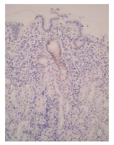
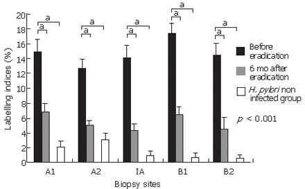
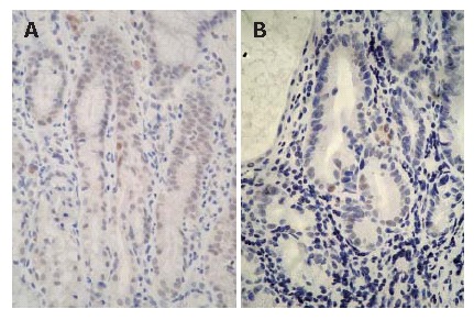
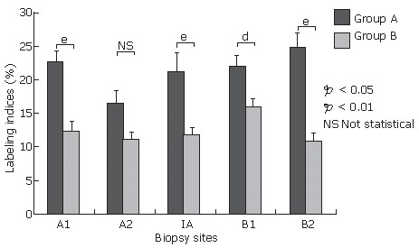
Results: p53 (DO-7 and PAb240) immunoreactivity was shown in the neck region of the gastric pits, however, quite a few cells were found to be immunopositive for p53 (PAb240) in the H pylori-infected gastric mucosa. The proportion of patients immunopositive for p53 (PAb240) was significantly reduced 6 mo after eradication [28/42 (66.7%) to 6/42 (14.3%)] (P < 0.05), while the biopsies taken from H pylori-negative patients showed no immunoreactivity for p53 (PAb240). p53 (PAb240)-positive patients were divided into two groups by the number of positive cells detected: one with more than six positive cells per 10 gastric pits (group A, n = 12), and the other with less than five positive cells per 10 gastric pits (group B, n = 30). Atrophy scores in group A were significant higher than those in group B at the greater curvature of the antrum (group A: 2.00 +/- 0.14 vs group B: 1.40 +/- 0.15, P = 0.012), the lesser curvature of the corpus (group A: 2.00 +/- 0.21 vs group B: 1.07 +/- 0.23, P = 0.017), and the greater curvature of the corpus (group A: 1.20 +/- 0.30 vs group B: 0.47 +/- 0.21, P = 0.031). Group A showed significant higher intestinal metaplasia scores than group B only at the lesser curvature of the antrum (group A: 2.10 +/- 0.41 vs group B: 1.12 +/- 0.29, P = 0.035).
Conclusion: H pylori-associated chronic gastritis expressed the mutant-type p53, which was significantly associated with more severe atrophic and metaplastic changes. H pylori eradication led to a significant reduction in the expression of the mutant-type p53. It is considered that H pylori-infected chronic gastritis is associated with a genetic instability that leads to gastric carcinogenesis, and H pylori eradication may prevent gastric cancer.
Figures
Similar articles
-
[The assessment of nitric oxide metabolites in gastric juice in Helicobacter pylori infected subjects in compliance with grade of inflammatory lesions in gastric mucosa].Pol Merkur Lekarski. 2008 Feb;24(140):95-100. Pol Merkur Lekarski. 2008. PMID: 18634262 Polish.
-
Effect of Helicobacter pylori infection and its eradication on cell proliferation, DNA status, and oncogene expression in patients with chronic gastritis.Gut. 1999 Jun;44(6):789-99. doi: 10.1136/gut.44.6.789. Gut. 1999. PMID: 10323879 Free PMC article.
-
Helicobacter pylori infection affects the expression of PCNA, p53, c-erbB-2 and Bcl-2 in the human gastric mucosa.Rev Esp Enferm Dig. 2003 Feb;95(2):97-104, 89-96. Rev Esp Enferm Dig. 2003. PMID: 12760717 English, Spanish.
-
Apoptosis in gastric epithelium induced by Helicobacter pylori infection: implications in gastric carcinogenesis.Am J Gastroenterol. 2001 Jan;96(1):16-26. doi: 10.1111/j.1572-0241.2001.03447.x. Am J Gastroenterol. 2001. PMID: 11197247 Review.
-
[The effect of Helicobacter pylori eradication on chronic gastritis].Nihon Rinsho. 2013 Aug;71(8):1442-8. Nihon Rinsho. 2013. PMID: 23967677 Review. Japanese.
Cited by
-
p53 inhibition of AP1-dependent TFF2 expression induces apoptosis and inhibits cell migration in gastric cancer cells.Am J Physiol Gastrointest Liver Physiol. 2009 Aug;297(2):G385-96. doi: 10.1152/ajpgi.90620.2008. Epub 2009 Jun 18. Am J Physiol Gastrointest Liver Physiol. 2009. PMID: 19541923 Free PMC article.
-
Helicobacter pylori Infection Activates the Akt-Mdm2-p53 Signaling Pathway in Gastric Epithelial Cells.Dig Dis Sci. 2015 Apr;60(4):876-86. doi: 10.1007/s10620-014-3470-2. Epub 2014 Dec 6. Dig Dis Sci. 2015. PMID: 25480405
-
Pseudo-mutant P53 is a unique phenotype of DNMT3A-mutated pre-leukemia.Haematologica. 2022 Nov 1;107(11):2548-2561. doi: 10.3324/haematol.2021.280329. Haematologica. 2022. PMID: 35199506 Free PMC article.
-
Ten-year prospective follow-up of histological changes at five points on the gastric mucosa as recommended by the updated Sydney system after Helicobacter pylori eradication.J Gastroenterol. 2012 Apr;47(4):394-403. doi: 10.1007/s00535-011-0504-9. Epub 2011 Dec 6. J Gastroenterol. 2012. PMID: 22138891
-
Immunohistochemical differences in gastric mucosal damage between nodular and non-nodular gastritis caused by Helicobacter pylori infection.J Clin Biochem Nutr. 2021 Sep;69(2):216-221. doi: 10.3164/jcbn.20-179. Epub 2021 Mar 25. J Clin Biochem Nutr. 2021. PMID: 34616112 Free PMC article.
References
-
- Marshall BJ, Armstrong JA, McGechie DB, Glancy RJ. Attempt to fulfil Koch's postulates for pyloric Campylobacter. Med J Aust. 1985;142:436–439. - PubMed
-
- Morris A, Nicholson G. Ingestion of Campylobacter pyloridis causes gastritis and raised fasting gastric pH. Am J Gastroenterol. 1987;82:192–199. - PubMed
-
- Fujioka T, Kubota T, Shuto R, Kodama R, Murakami K, Perparim K, Nasu M. Establishment of an animal model for chronic gastritis with Helicobacter pylori: potential model for long-term observations. Eur J Gastroenterol Hepatol. 1994;6 Suppl 1:S73–S78. - PubMed
-
- Kuipers EJ, Uyterlinde AM, Peña AS, Roosendaal R, Pals G, Nelis GF, Festen HP, Meuwissen SG. Long-term sequelae of Helicobacter pylori gastritis. Lancet. 1995;345:1525–1528. - PubMed
-
- Sakaki N, Momma K, Egawa N, Yamada Y, Kan T, Ishiwata J. The influence of Helicobacter pylori infection on the progression of gastric mucosal atrophy and occurrence of gastric cancer. Eur J Gastroenterol Hepatol. 1995;7 Suppl 1:S59–S62. - PubMed
Publication types
MeSH terms
Substances
LinkOut - more resources
Full Text Sources
Research Materials
Miscellaneous
