. 2007 Aug 1;405(3):569-81.
doi: 10.1042/BJ20061812.

Cdc34 C-terminal tail phosphorylation regulates Skp1/cullin/F-box (SCF)-mediated ubiquitination and cell cycle progression

Affiliations

Cdc34 C-terminal tail phosphorylation regulates Skp1/cullin/F-box (SCF)-mediated ubiquitination and cell cycle progression

Martin Sadowski et al. Biochem J. .

Abstract

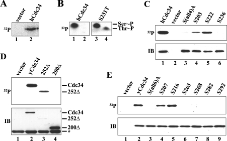
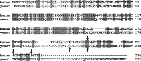
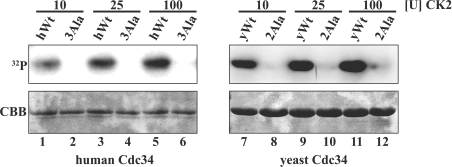
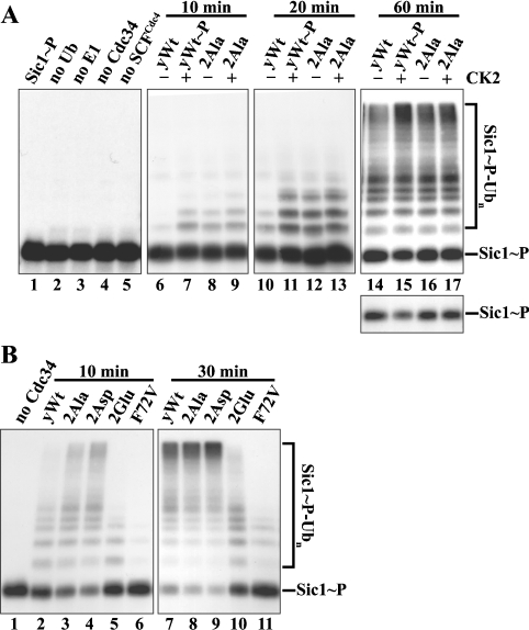
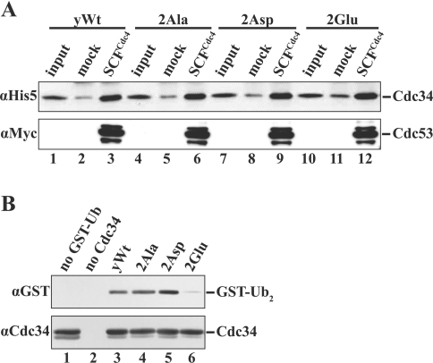
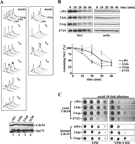
The ubiquitin-conjugating enzyme Cdc34 (cell division cycle 34) plays an essential role in promoting the G1-S-phase transition of the eukaryotic cell cycle and is phosphorylated in vivo. In the present study, we investigated if phosphorylation regulates Cdc34 function. We mapped the in vivo phosphorylation sites on budding yeast Cdc34 (yCdc34; Ser207 and Ser216) and human Cdc34 (hCdc34 Ser203, Ser222 and Ser231) to serine residues in the acidic tail domain, a region that is critical for Cdc34's cell cycle function. CK2 (protein kinase CK2) phosphorylates both yCdc34 and hCdc34 on these sites in vitro. CK2-mediated phosphorylation increased yCdc34 ubiquitination activity towards the yeast Saccharomyces cerevisiae Sic1 in vitro, when assayed in the presence of its cognate SCFCdc4 E3 ligase [where SCF is Skp1 (S-phase kinase-associated protein 1)/cullin/F-box]. Similarly, mutation of the yCdc34 phosphorylation sites to alanine, aspartate or glutamate residues altered Cdc34-SCFCdc4-mediated Sic1 ubiquitination activity. Similar results were obtained when yCdc34's ubiquitination activity was assayed in the absence of SCFCdc4, indicating that phosphorylation regulates the intrinsic catalytic activity of Cdc34. To evaluate the in vivo consequences of altered Cdc34 activity, wild-type yCdc34 and the phosphosite mutants were introduced into an S. cerevisiae cdc34 deletion strain and, following synchronization in G1-phase, progression through the cell cycle was monitored. Consistent with the increased ubiquitination activity in vitro, cells expressing the phosphosite mutants with higher catalytic activity exhibited accelerated cell cycle progression and Sic1 degradation. These studies demonstrate that CK2-mediated phosphorylation of Cdc34 on the acidic tail domain stimulates Cdc34-SCFCdc4 ubiquitination activity and cell cycle progression.


Figures

Figure 1
Figure 1. Identification of the in vivo phosphorylation sites on hCdc34 and yCdc34
(A) HEK-293 cells were transiently transfected with either pCMV-Tag2 (vector) or pCMV-Tag2-hCdc34(Wt) and labelled with [32P]Pi. Human Cdc34 was immunoprecipitated, separated by SDS/PAGE and visualized by autoradiography. (B) Wild-type 32P-labelled hCdc34 (left panel) or its mutant derivative S231T (right panel) were subjected to phospho amino acid analysis. The positions of phosphoserine (Ser∼P) and phosphothreonine (Thr∼P) are indicated. (C) HEK-293 cells were transiently transfected with either pCMV-Tag2 (vector) or pCMV-Tag2 constructs expressing wild-type hCdc34 or its mutant derivatives S(all4)A (S203A/S222A/S231A/S236A), S203 (S222A/S231A/S236A), S222 (S203A/S231A/S236A) and S236 (S203A/S222A/S231A) and subjected to the same treatment as in (A). After SDS/PAGE, proteins were visualized by autoradiography (32P) or by immunoblotting (IB) with an anti-hCdc34 antibody. (D) Yeast cells (W303-1A) were transformed with empty pESC-TRP (vector) or pESC1 constructs expressing either yeast wild-type Cdc34 (yCdc34) or its C-terminal truncation derivatives 252Δ (residues 1–252) and 200Δ (residues 1–200). After labelling with [32P]Pi, cells were lysed, and yCdc34 was immunoprecipitated, separated by SDS/PAGE and visualized by autoradiography (32P) or by immunoblotting (IB) with anti-FLAG antibody. The asterisk indicates the position of the light immunoglobulin chain of the anti-FLAG beads. (E) Yeast cells (W303-1A) were transformed with empty pESC-TRP (vector) or pESC1 constructs expressing either yeast wild-type Cdc34 (yCdc34) or its mutant derivatives S(all6)A (S207A/S216A/S263A/S268A/S282A/S292A), S207 (S216A/S263A/S268A/S282A/S292A), S216 (S207A/S263A/S268A/S282A/S292A), S263 (S207A/S216A/S268A/S282A/S292A), S268 (S207A/S216A/S263A/S282A/S292A), S282 (S207A/S216A/S263A/S268A/S292A) and S292 (S207A/S216A/S263A/S268A/S282A). Cells were subjected to metabolic 32P labelling as in (D), and proteins were visualized by autoradiography (32P) or by immunoblotting (IB) with an anti-Cdc34 antibody.
Figure 2
Figure 2. Sequence alignment of yCdc34 and hCdc34
The amino acid sequences of hCdc34 and yeast Cdc34 were aligned using ClustalW (1.82). Identical residues are shaded in dark grey, conserved residues in light grey, and the in vivo phosphorylation sites are marked by arrows.
Figure 3
Figure 3. CK2 phosphorylates the in vivo phosphorylation sites of hCdc34 and yCdc34
Purified recombinant human wild-type (hWt, left-hand panel) and yeast wild-type Cdc34 (yWt, right-hand panel) and their respective alanine phosphorylation site mutants [3Ala (S203A/S222A/S231A) and 2Ala (S207A/S216A)] were incubated with increasing amounts of CK2 in the presence of [γ-32P]ATP in vitro. Proteins were separated by SDS/PAGE and visualized by autoradiography (32P) and by Coomassie staining (CBB).
Figure 4
Figure 4. The phosphorylation sites of yeast Cdc34 are important for Sic1 ubiquitination in vitro
(A) Phosphorylation of recombinant yCdc34 by CK2 enhances SCFCdc4-mediated ubiquitination of Sic1. To monitor Sic1 ubiquitination, SCFCdc4 complexes were supplemented with E1, ubiquitin, ATP, phosphorylated, 32P-labelled Sic1 (lane 1), and yeast wild-type Cdc34 (yWt), which was pre-incubated in the absence (–) or presence (+) of CK2. Prior to ubiquitination, CK2 was inhibited by 2 μM heparin. As controls, the phosphosite mutant 2Ala was subjected to the same treatment (lanes 8, 9, 12, 13, 16 and 17) and single components were omitted from the reaction (lanes 2–5). After incubation for 10, 20 or 60 min at 26 °C, proteins were separated by SDS/PAGE and visualized by autoradiography. The lower panel shows a shorter exposure of the unconjugated Sic1-P substrate remaining. (B) Mutation of the phosphorylation sites of recombinant yCdc34 alters SCFCdc4-mediated ubiquitination of Sic1. Yeast wild-type Cdc34 (yWt; lanes 2 and 7), the phosphosite mutants 2Ala (lanes 3 and 8), 2Asp (lanes 4 and 9), 2Glu (lanes 5 and 10) or the point mutant yCdc34(F72V) (lanes 6 and 11) were assayed as in (A), except that incubation times were 10 and 30 min. As a control, yCdc34 was omitted from the reaction (lane 1).
Figure 5
Figure 5. The phosphorylation sites of yeast Cdc34 regulate ubiquitin-conjugating activity independent of SCF E3 ligase in vitro
(A) Mutation of the phosphorylation sites of recombinant yeast Cdc34 do not affect binding to SCFCdc4 in vitro. Protein-binding studies of yeast wild-type Cdc34 (yWt; lanes 1–3) and the phosphosite mutants 2Ala (lanes 4–6), 2Asp (lanes 7–9) and 2Glu (lanes 10–12) with either SCFCdc4 or mock-treated anti-FLAG beads were performed as described in the Materials and methods section. After extensive washing of the beads, proteins were separated by SDS/PAGE, and His6-tagged yCdc34 was visualized by immunoblotting with anti-His5 antibody (upper panel). As a control for SCFCdc4 loading, blots were reprobed with anti-Myc antibody to detect Myc-tagged Cdc53 (lower panel). (B) The catalytic activity of recombinant yeast wild-type Cdc34 (yWt; lane 3) and the phosphosite mutants 2Ala (lane 4), 2Asp (lane 5) and 2Glu (lane 6) was compared by monitoring the formation of mono-ubiquitinated GST–ubiquitin (GST–Ub2) in the absence of SCF E3 ligase in vitro, as described in the Materials and methods section. As controls, GST–Ub (lane 1) and Cdc34 (lane 2) were omitted from the reaction. Proteins were separated by SDS/PAGE and visualized by immunoblotting with antibodies directed against the GST tag (αGST) present on GST–Ub and yCdc34 (αCdc34) respectively.
Figure 6
Figure 6. Mutation of the yCdc34 phosphorylation sites affects cell cycle progression and Sic1 turnover in vivo
(A) Exponentially, asynchronously (async) growing yeast cells (YMS034) expressing wild-type yCdc34 (yWt) or the phosphosite mutants 2Ala, 2Asp (left-hand panel) and 2Glu (right-hand panel) were synchronized in G1-phase by α-factor arrest, released into the cell cycle by incubating in fresh medium, and aliquots were withdrawn at the time points indicated (t0=0 min). Cellular DNA was stained with propidium iodide and analysed by flow cytometry. The histograms are representative of at least three independent experiments. Equivalent expression levels of wild-type (lane 1) and mutant yCdc34s (lanes 2–4) were confirmed by immunoblotting of protein lysates of cells with anti-Cdc34 antibody (upper panel) and even loading was confirmed by immunoblotting with anti-Spc72 antibody (lower panel). (B) Yeast strains expressing either wild-type yCdc34 (yWt), 2Ala, 2Asp or F72V were synchronized in G1 of the cell cycle by α-factor arrest. At the indicated times after release, cell extracts were prepared and analysed by immunoblotting with Sic1 antibody (left-hand panel) and actin antibody as a loading control (right-hand panel). Sic1 levels of cells expressing wild-type yCdc34 (solid black line), F72V (dotted grey line) and the phosphosite mutants 2Ala (dashed grey line) and 2Asp (dotted black line) of three independent experiments were quantified (lower panel). The data points represent the means±S.D. (n=3). (C) Mutation of the phosphorylation sites confers increased resistance on cycloheximide (CHX). Serial 10-fold dilutions of yeast cells (YES71) containing either yeast wild-type Cdc34 (yWt) or the phosphosite mutants 2Ala, 2Asp and 2Glu or the point mutant yCdc34(F72V) were spotted on to YPD plates containing no (upper left panel) or 1.25 μg/ml CHX (upper right panel) and then incubated at 30 °C. Yeast cells containing human wild-type Cdc34 (hWt) or the phosphosite mutants 3Ala and 3Asp were subjected to the same analysis (lower panels) except that 0.75 μg/ml CHX was employed.

Similar articles

Cited by

References

    1. Pickart C., Eddins M. Ubiquitin: structures, functions, mechanisms. Biochim. Biophys. Acta. 2004;1695:55–72. - PubMed
    1. Thrower J. S., Hoffman L., Rechsteiner M., Pickart C. M. Recognition of the polyubiquitin proteolytic signal. EMBO J. 2000;19:94–102. - PMC - PubMed
    1. Sun L., Chen Z. J. The novel functions of ubiquitination in signaling. Curr. Opin. Cell Biol. 2004;16:119–126. - PubMed
    1. Pickart C. M., Fushman D. Polyubiquitin chains: polymeric protein signals. Curr. Opin. Chem. Biol. 2004;8:610–616. - PubMed
    1. Petroski M., Deshaies R. Function and regulation of cullin–RING ubiquitin ligases. Nat. Rev. Mol. Cell Biol. 2005;6:8–20. - PubMed

Publication types

MeSH terms