. 2007 May;64(3):738-54.
doi: 10.1111/j.1365-2958.2007.05688.x.

An antisense RNA controls synthesis of an SOS-induced toxin evolved from an antitoxin

Affiliations

An antisense RNA controls synthesis of an SOS-induced toxin evolved from an antitoxin

Mitsuoki Kawano et al. Mol Microbiol. 2007 May.

Abstract

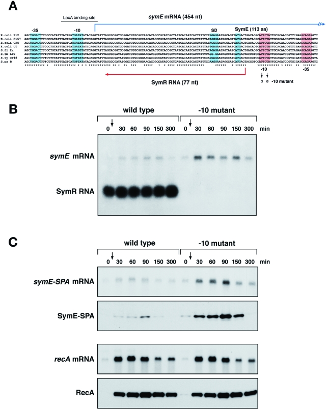
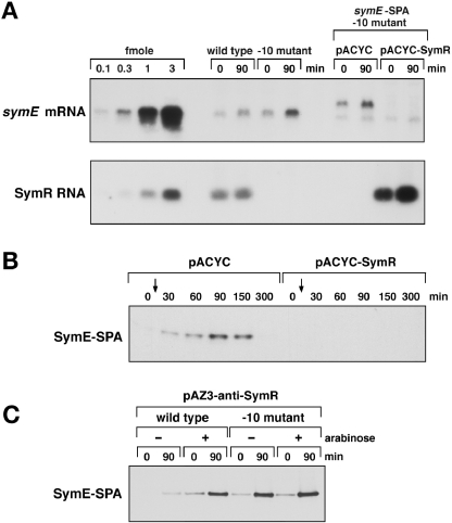
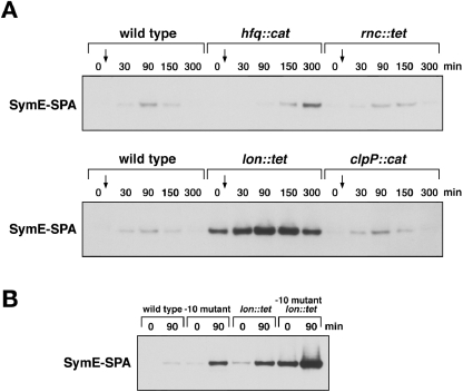
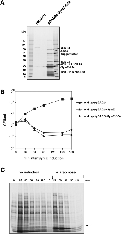
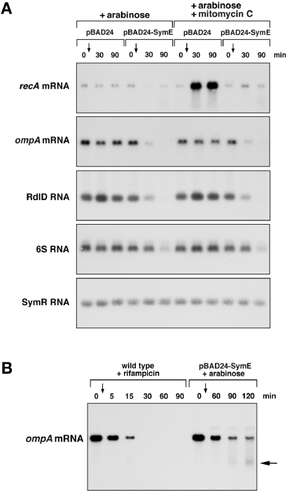
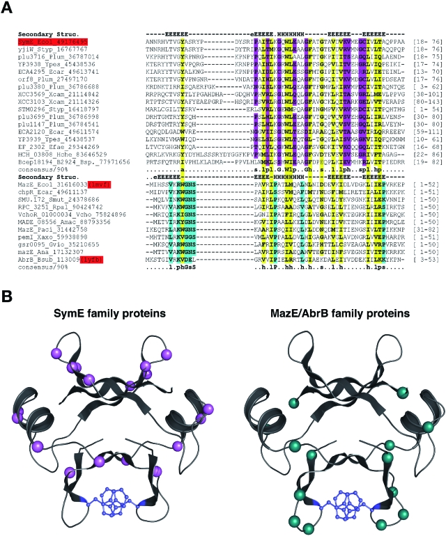
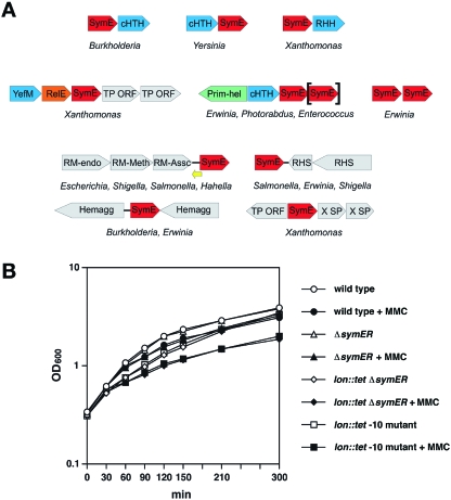
Only few small, regulatory RNAs encoded opposite another gene have been identified in bacteria. Here, we report the characterization of a locus where a small RNA (SymR) is encoded in cis to an SOS-induced gene whose product shows homology to the antitoxin MazE (SymE). Synthesis of the SymE protein is tightly repressed at multiple levels by the LexA repressor, the SymR RNA and the Lon protease. SymE co-purifies with ribosomes and overproduction of the protein leads to cell growth inhibition, decreased protein synthesis and increased RNA degradation. These properties are shared with several RNA endonuclease toxins of the toxin-antitoxin modules, and we show that the SymE protein represents evolution of a toxin from the AbrB fold, whose representatives are typically antitoxins. We suggest that SymE promotion of RNA cleavage may be important for the recycling of RNAs damaged under SOS-inducing conditions.


Figures

Fig. 1
Fig. 1
SymR RNA represses SymE translation. A. Genetic organization of the symER locus. Multiple sequence alignments of the different symER sequences were constructed using clustalw. The species correspond to E. coli strains K12, O157:H7, CFT073 and UT189, Shigella flexneri 2a, Salmonella typhimurium LT2, S. typhi CT18 and S. paratyphi B. The −10 and −35 promoter sequences, the Shine-Dalgarno sequence and initiation codon of symE are indicated by blue boxes. The −10 and −35 sequences of symR are indicated by red boxes. B. symE mRNA and SymR RNA levels in MG1655 and the −10 symR promoter mutant. Total RNA was isolated from wild-type MG1655 and −10 mutant strains grown in LB medium at 37°C at 0, 30, 60, 90, 150 and 300 min after treatment with 1 μg ml−1 mitomycin C. Samples (5 μg) were analysed by Northern hybridization using oligonucleotide probes specific to symE and SymR. C. symE-SPA mRNA and protein and recA mRNA and protein levels in MG1655 symE-SPA and the −10 symR promoter mutant. Total RNA and cell lysates were prepared from MG1655 symE-SPA and symE-SPA −10 mutant strains in grown in LB medium at 37°C at 0, 30, 60, 90, 150 and 300 min after treatment with 1 μg ml−1 mitomycin C. RNA samples (5 μg) were analysed by Northern hybridization using oligonucleotide probes specific to symE and recA, and cell lysates were analysed by immunoblot assays using monoclonal anti-FLAG M2-AP and polyclonal anti-RecA antibodies.
Fig. 2
Fig. 2
Effects of different SymR RNA levels. A. Quantitative Northern analysis of symE and SymR RNA levels. Total RNA (10 μg) isolated for the Northern analysis in Fig. 1B and total RNA (0.5 μg) isolated from the samples used for the Western analysis in Fig. 2B was separated alongside in vitro synthesized RNA (0.1, 0.3, 1 and 3 fmol) on 1.2% agarose gels. The cellular levels of symE mRNA and SymR RNA were determined from the ratio of the signals of control RNAs to the cellular RNAs. B. SymR RNA expressed in trans can repress SymE synthesis. The symE-SPA −10 mutant carrying pACYC and pACYC-SymR was grown to OD600∼0.3 in LB medium containing tetracycline at 37°C. Cell lysates prepared at 0, 30, 60, 90, 150 and 300 min after treatment with 1 μg ml−1 mitomycin C were analysed by immunoblot assays using monoclonal anti-FLAG M2-AP antibodies. C. Expression of an anti-antisense RNA leads to increased SymE-SPA synthesis. MG1655 PCP18-araE symE-SPA and MG1655 PCP18-araE symE-SPA −10 mutant strains carrying pAZ3-anti-SymR were grown to OD600∼0.2 in LB medium containing chloramphenicol at 37°C. The cultures were split and half of each culture was treated with 0.02% arabinose for 30 min (0 min). All cultures were then treated with 1 μg ml−1 mitomycin C for 90 min (90 min). Cell extracts prepared at the 0 and 90 min time points were analysed by immunoblot assays using monoclonal anti-FLAG M2-AP antibodies.
Fig. 3
Fig. 3
SymE protein is degraded by the Lon protease. A. SymE-SPA synthesis in hfq, rnc, lon and clpP mutant strains. The MG1655 symE-SPA strain and the corresponding mutant derivatives were grown to OD600∼0.3 in LB medium at 37°C. Cell lysates prepared at 0, 30, 90, 150 and 300 min after treatment with 1 μg ml−1 mitomycin C were analysed by Western hybridization using monoclonal anti-FLAG M2-AP antibodies. B. Relative SymE-SPA levels in the −10 symR promoter and lon mutant backgrounds. The MG1655 symE-SPA, symE-SPA −10 mutant, symE-SPA lon mutant, symE-SPA −10 lon mutant strains were grown to OD600∼0.5 in LB medium at 37°C. Cell lysates prepared at 0 and 90 min after treatment with 1 μg ml−1 mitomycin C were analysed by immunoblot assays using monoclonal anti-FLAG M2-AP antibodies.
Fig. 4
Fig. 4
Overexpression of ribosome-associated SymE leads to reduced colony formation and decreased protein synthesis. A. Purification of SymE-SPA. Extracts prepared from MG1655 kan-PCP18-araE carrying either pBAD24 or pBAD-SymE-SPA were subjected to affinity purification as described in Experimental procedures. The samples then were analysed by 4–20% SDS-PAGE and visualized using GelCode Blue Stain Reagent. The identities of the indicated protein bands were determined by LC/MS/MS mass spectrometry. B. SymE overexpression results in reduced colony-forming ability. MG1655 PCP18-araE carrying pBAD24, pBAD-SymE and pBAD-SymE-SPA were grown to OD600∼0.3 at 37°C in LB medium containing ampicillin. Two min after time 0, 0.02% arabinose was added to induce transcription of symE or symE-SPA. At the indicated time points, cells were diluted and plated on LB solid medium containing ampicillin. C. SymE overexpression results in reduced protein synthesis. SDS-PAGE analysis of in vivo total protein synthesis after the induction of SymE. GSO120/pBAD-SymE cells were grown in M9 medium with glycerol, casamino acids and ampicillin with or without arabinose. Aliquots (0.5 ml) of the cell culture were taken at the indicated time points and added to 20 μCi of Tran35S-label containing 35S-methionine and 35S-cysteine as described in Experimental procedures. The band indicated with an arrow is SymE (∼12 kDa). This experiment was performed multiple times. The figure is a representative experiment.
Fig. 5
Fig. 5
SymE exhibits ribonuclease activity. A. SymE overexpression leads to reduced levels of some RNAs. MG1655 PCP18-araE carrying either pBAD24 or pBAD-SymE were grown to OD600∼0.5 at 37°C in M9 minimal medium supplemented with glycerol and casamino acids. Total RNA isolated at 0, 30, and 90 min after treatment with 0.02% arabinose and 0.02% arabinose plus 1 μg ml−1 mitomycin C. Samples (5 μg) were analysed by Northern hybridization using oligonucleotide probes specific to recA, ompA, RdlD, 6S and SymR. B. Detection of degradation products upon SymE overexpression. MG1655 cells were grown to OD600∼0.5 at 37°C in LB medium, and total RNA was isolated at 0, 5, 15, 30, 60 and 90 min after treatment with 300 μg ml−1 rifampicin. MG1655 PCP18-araE cells carrying pBAD-SymE were grown to OD600∼0.25 at 37°C in LB medium, and total RNA was isolated at 0, 60, 90, and 120 min after treatment with 0.02% arabinose. Samples (5 μg) were analysed by Northern hybridization using an oligonucleotide probe specific to ompA. Degradation products are indicated by the larger arrow.
Fig. 6
Fig. 6
SymE is an AbrB superfamily member that has acquired a toxin-like function. A. Multiple alignment of the SymE family and other members of the AbrB superfamily. Proteins are denoted by gene name, species abbreviation, and GenBank Identifier (gi) number; separated by underscores. Positions strongly conserved at or above the 90% applied on the entire family of proteins only among the classic SymE family proteins are shaded pink, whereas those similarly conserved only in the classic MazE/AbrB family are shaded aqua and those conserved between both are shaded yellow. Consensus similarity designations are as follows: h, hydrophobic residues (ACFILMVWY); s, small residues (AGSVCDN); p, polar residues. Secondary structure assignments obtained from the jpred prediction for the SymE family and from the crystal structures (like PDB: 1mvf and 1ub4) for the rest of the AbrB superfamily are shown above the alignment where E represents a strand and H represents a helix. The region shown in the alignment spans the entire length of the DNA binding domain of the classical AbrB proteins. The boundaries are shown to the right. Species abbreviations are as given for the full alignment in Fig. S2. B. Models of the classic SymE family and classic MazE/AbrB family proteins. An idealized version of the AbrB fold was constructed using the consensus sequence derived from the hidden Markov model for the entire fold using SWISSMODEL server of SWISSPDB and the 1mvf structure as a template; in both cases the structure is depicted as a dimer formed by two interlocking monomers, which was achieved using the oligomer mode in SWISSMODEL (Guex and Peitsch, 1997). The positions that are conserved in the SymE family at the 90% consensus are coloured pink as in Fig. 6A. The majority of them line a groove on one face of the protein. This surface faces away from the surface that contains the most conserved positions unique to the classical members of the MazE/AbrB superfamily (coloured aqua). The uniquely conserved regions include polar residues that could potentially mediate the key interactions of SymE with the ribosome or target RNA. The aromatic residues involved in stabilizing the dimer through a π–π stacking interaction are shown in blue.
Fig. 7
Fig. 7
Physiological role of SymE. A. Gene neighbourhoods and predicted operons of the SymE family of genes. The orientation of the genes is denoted by the direction of the arrow. The yellow arrow denotes the SymR RNA that is transcribed in the SymE proper group. The genes are all labelled as per the gene products: SymE = SymE family; cHTH = a Cro/cI type transcriptional regulator, RHH = a MetJ/Arc-type transcription factor, YefM = a YefM-type transcription factor, RelE = a toxin homologous to the RelE toxins, Prim-Hel = the gene encoding a two module protein with a N-terminal DNAG-type primase domain and C-terminal MCM-like AAA + helicase domain. Shown in grey are other genes belonging to the larger genomic context, in the mobile islands in which the SymE family genes are inserted. These include transposons = TP ORF and restriction-modification operons = RM-endo, RM-Meth and RM-Assc for the endonuclease, methylase and accessory subunits respectively. Representative examples of neighbourhoods with the Xanthomonas-specific secreted protein (X SP) and the haemagglutinin and RHS cell-surface complex genes are also shown. The SymE enclosed in ‘[]’indicates the presence of an optional second SymE gene in some of these neighbourhoods. B. ΔsymER growth after DNA damage. Wild type, ΔsymER, lon::tetΔsymER and lon::tet−10 mutant cells were grown to OD600∼0.3 at 37°C in LB medium. Expression of SymE was induced by the addition of 1 μg ml−1 mitomycin C, and cell growth was monitored by measuring OD600 at the indicated time points.

Similar articles

Cited by

References

    1. Aizenman E, Engelberg-Kulka H, Glaser G. An Escherichia coli chromosomal ‘addiction module’ regulated by guanosine 3′,5′-bispyrophosphate: a model for programmed bacterial cell death. Proc Natl Acad Sci USA. 1996;93:6059–6063. - PMC - PubMed
    1. Altschul SF, Madden TL, Schaffer AA, Zhang J, Zhang Z, Miller W, Lipman DJ. Gapped blast and psi-blast: a new generation of protein database search programs. Nucleic Acids Res. 1997;25:3389–3402. - PMC - PubMed
    1. Anantharaman V, Aravind L. New connections in the prokaryotic toxin-antitoxin network: relationship with the eukaryotic nonsense-mediated RNA decay system. Genome Biol. 2003;4:R81. - PMC - PubMed
    1. Anantharaman V, Aravind L. Novel predicted RNAses with a PIN Domain-like Fold. RNA Biol. 2006;3:18–27. - PubMed
    1. Aravind L, Makarova KS, Koonin EV. Holliday junction resolvases and related nucleases: identification of new families, phyletic distribution and evolutionary trajectories. Nucleic Acids Res. 2000;28:3417–3432. - PMC - PubMed

Publication types

MeSH terms

LinkOut - more resources