. 2008 Jan;32(1):131-41.
doi: 10.1016/j.leukres.2007.03.025. Epub 2007 Apr 25.

Guanine nucleotide depletion inhibits pre-ribosomal RNA synthesis and causes nucleolar disruption

Affiliations

Guanine nucleotide depletion inhibits pre-ribosomal RNA synthesis and causes nucleolar disruption

Min Huang et al. Leuk Res. 2008 Jan.

Abstract

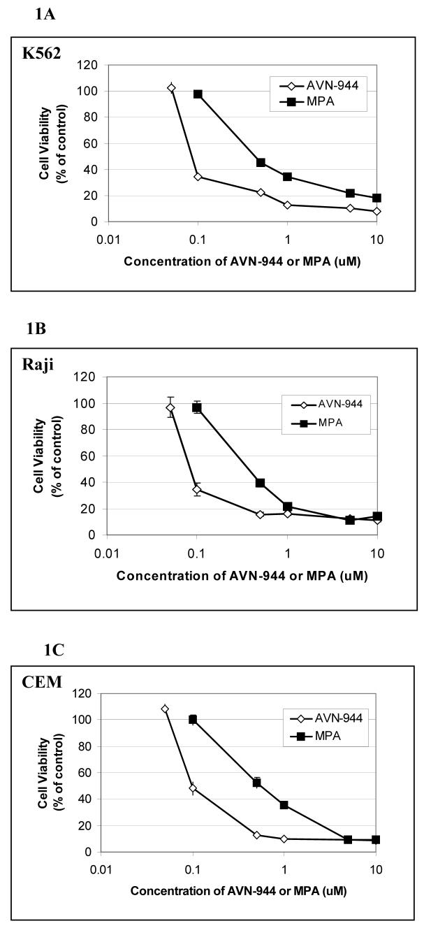
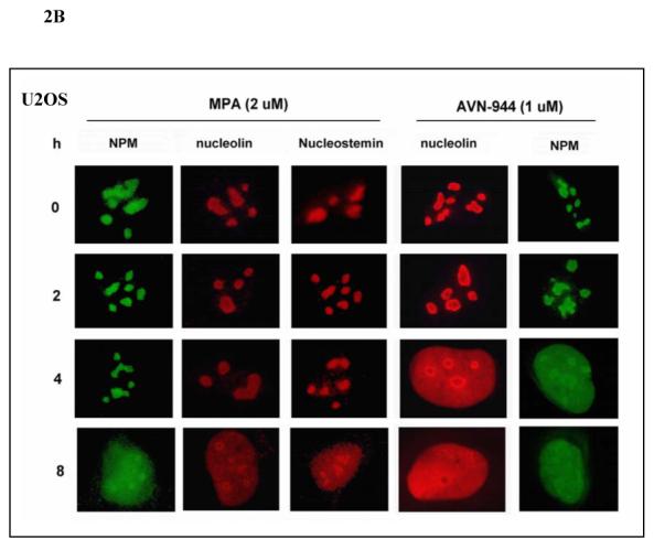
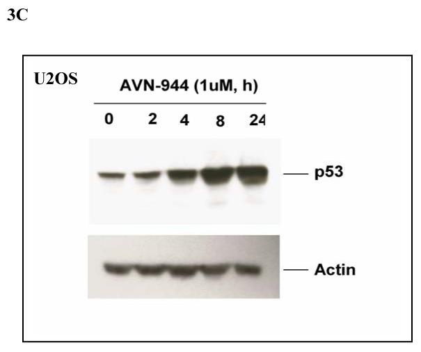
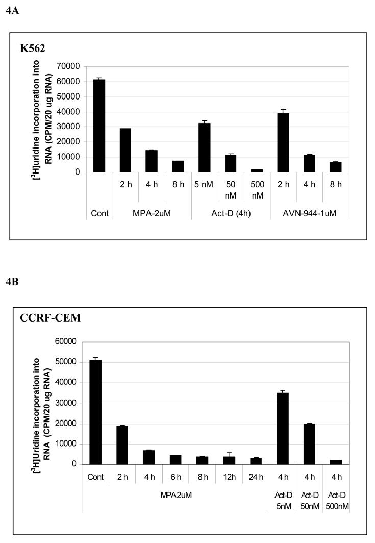
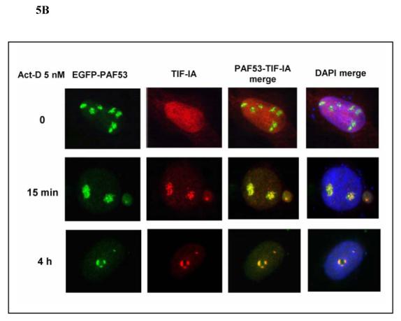
Inosine monophosphate dehydrogenase (IMPDH) is a pivotal enzyme in the de novo pathway of guanine nucleotide biosynthesis. Inhibitors of this enzyme decrease intracellular guanine nucleotide levels by 50-80% and have potential as anti-neoplastic agents. Both mycophenolic acid (MPA) and AVN-944 are highly specific inhibitors of IMPDH that cause cell cycle arrest or apoptosis in lymphocytes and leukemic cell lines. We have examined the mechanisms by which these two agents cause cytotoxicity. Both MPA and AVN-944 inhibit the growth of K562 cells, and induce apoptosis in Raji B and CCRF-CEM T cells. Both compounds strikingly inhibit RNA synthesis within 2 h of exposure. Depletion of guanine nucleotides by MPA and AVN-944 also causes an early and near-complete reduction in levels of the 45S precursor rRNA synthesis and the concomitant translocation of nucleolar proteins including nucleolin, nucleophosmin, and nucleostemin from the nucleolus to the nucleoplasm. This efflux correlates temporally with the sustained induction of p53 in cell lines with wild-type p53. We conclude that inhibition of IMPDH causes a primary reduction in rRNA synthesis and secondary nucleolar disruption and efflux of nucleolar proteins that most likely mediate cell cycle arrest or apoptosis. The ability of AVN-944 to induce apoptosis in a number of leukemic cell lines supports its potential utility in the treatment of hematologic malignancies.


Figures

Fig. 1
Fig. 1. Inhibition of cell growth by MPA and AVN-944 in human leukemia K562 cells, Raji, and CCRF-CEM cells
K562 (A), Raji (B), and CCRF-CEM (C) cells were exposed to 1-10 μM MPA or 1-10 μM AVN-944 for 96 h. Cytotoxicity assays were performed by the modified MTT method. All assays were performed in triplicate. All values represent the mean +/− SEM.
Fig. 2
Fig. 2. Effect of MPA and AVN-944 on the localization of nucleophosmin, nucleolin, and nucleostemin and reversibility by guanosine
(A) U2OS cells were treated with 2 μM MPA in the absence or presence of 100 μM guanosine for 24 h. (B) U2OS cells were treated with 2 μM MPA or 1 μM AVN-944 for 2 h, 4 h, and 8 h. Cells were then fixed, permeabilized, stained for nucleolin, nucleostemin, or NPM, as described in Materials and Methods, and observed using fluorescent microscopy.
Fig. 3
Fig. 3. Effects of AVN-944 on p53 and ARF
MCF-7 cells (A) or 293 T cells (B) were treated with AVN-944 at the time and concentration indicated. Cells were then fixed, permeabilized, and stained for p53 and ARF as described in the Materials and Methods. (C) U2OS cells were treated with AVN-944 at the time and concentration indicated, and lysed as described in Materials and Methods. 30 μg of total protein were loaded for immunoblot analysis for p53.
Fig. 4
Fig. 4. Effects of MPA, AVN-944, and Actinomycin D on total RNA synthesis and prerRNA synthesis in K562 and CCRF-CEM cells
K562 cells were treated with MPA, AVN-944, or actinomycin-D at the concentrations and times indicated and [3H]-uridine incorporation into RNA was measured (A, B); the effect of MPA on the expression of pre-rRNA levels was determined by RT-PCR (C) as described in Materials and Methods. Primers amplified the 394 bp fragment extending from the 3' end of 18 S rRNA to the internal transcribed sequence (ITS) located between 18 S rRNA and 5.8 S rRNA of the intact pre-rRNA (shown as model figure, C). Analysis of rRNA processing (D). Extracted total [3H]-uridine-labeled RNAs from untreated K562 cells and cells treated with 2 μM MPA or 1 μM AVN-944 were separated on 1% agarose gel containing formaldehyde, transferred to membrane, and detected by autoradiography as described in Materials and Methods. The bottom panel shows 28S and 18S ribosomal RNA after transfer.
Fig. 5
Fig. 5. MPA and actinomycin-D induce a distinct co-translocation of EGFP-tagged PAF53 (Pol I subunit) and myc-tagged TIF-IA to the nucleolar cap
PAF53 cDNA subcloned into EGFP-C3 and TIF-IA cDNA subcloned into pcDNA3 were transfected into U2OS cells. 24 h after transfection, cells were treated with MPA (A) or actinomycin-D (B) at the concentrations and for the times indicated. The myc-tagged TIF-IA was fixed, permealized, and stained as described in the Materials and Methods, and observed using fluorescent microscopy.
Fig. 6
Fig. 6. Actinomycin-D induces translocation of NPM, nucleolin, and nucleostemin
U2OS cells were treated with actinomycin-D at 5 nM and 500 nM. Cells were then fixed, permealized, and stained for NPM, nucleolin, and nucleostemin as described in the Materials and Methods, and observed using fluorescent microscopy.

References

    1. Zimmermann AG, Gu JJ, Laliberte J, et al. Inosine-5′-monophosphate dehydrogenase: regulation of expression and role in cellular proliferation and T lymphocyte activation. Prog Nucleic Acid Res Mol Biol. 1998;61:181–209. - PubMed
    1. Allison AC, Eugui EM. Mechanisms of action of mycophenolate mofetil in preventing acute and chronic allograft rejection. Transplantation. 2005;80:S181–190. - PubMed
    1. Laliberte J, Yee A, Xiong Y, et al. Effects of guanine nucleotide depletion on cell cycle progression in human T lymphocytes. Blood. 1998;91:2896–2904. - PubMed
    1. Heinschink A, Raab M, Daxecker H, et al. In vitro effects of mycophenolic acid on cell cycle and activation of human lymphocytes. Clin Chim Acta. 2000;300:23–28. - PubMed
    1. Yalowitz JA, Pankiewicz K, Patterson SE, et al. Cytotoxicity and cellular differentiation activity of methylenebis(phosphonate) analogs of tiazofurin and mycophenolic acid adenine dinucleotide in human cancer cell lines. Cancer Lett. 2002;181:31–38. - PubMed

Publication types