. 2007 Jun 15;282(24):17613-22.
doi: 10.1074/jbc.M702799200. Epub 2007 Apr 26.

Phosphorylation of human CTP synthetase 1 by protein kinase C: identification of Ser(462) and Thr(455) as major sites of phosphorylation

Affiliations

Phosphorylation of human CTP synthetase 1 by protein kinase C: identification of Ser(462) and Thr(455) as major sites of phosphorylation

Yu-Fang Chang et al. J Biol Chem. .

Abstract

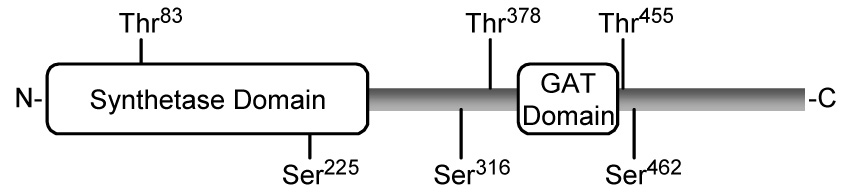
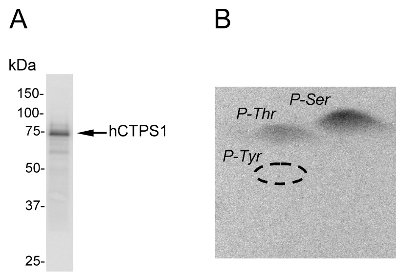
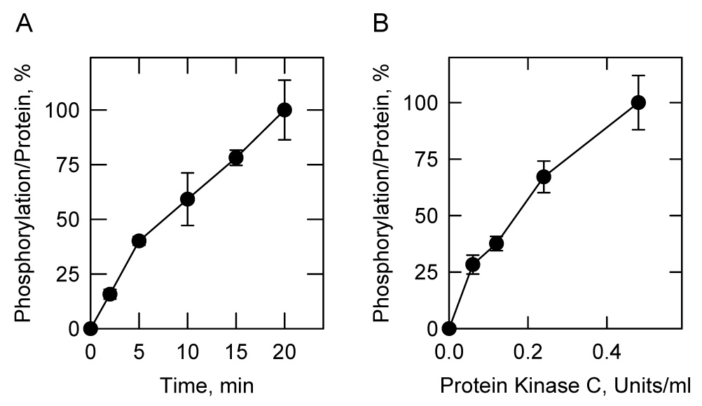
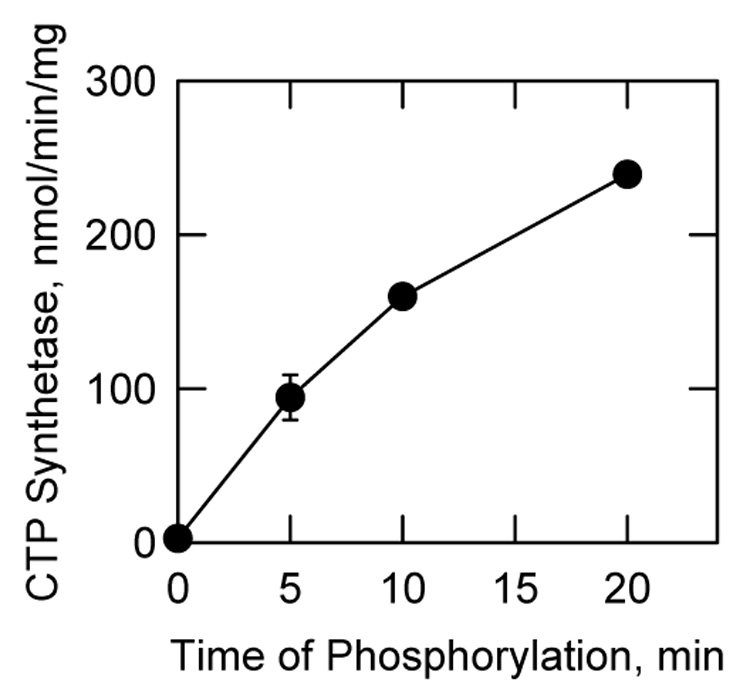
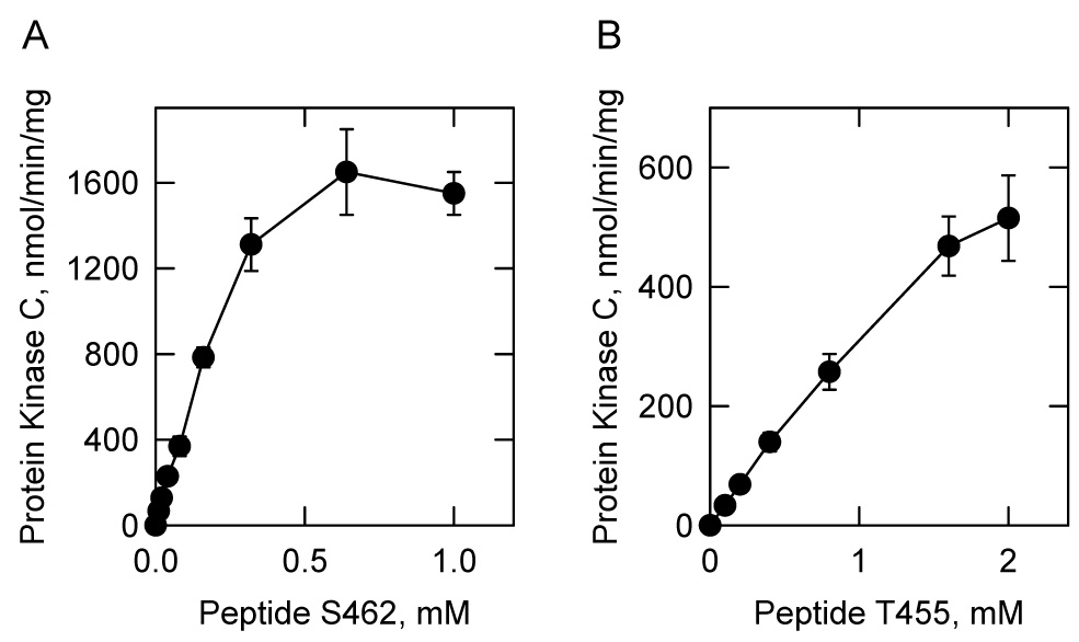
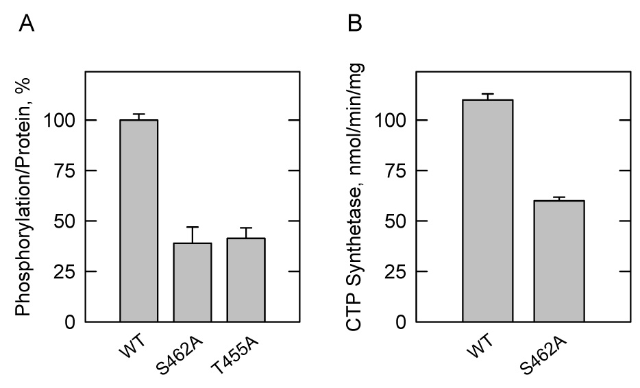
Phosphorylation of human CTP synthetase 1 by mammalian protein kinase C was examined. Using purified Escherichia coli-expressed CTP synthetase 1 as a substrate, protein kinase C activity was time- and dose-dependent and dependent on the concentrations of ATP and CTP synthetase 1. The protein kinase C phosphorylation of the recombinant enzyme was accompanied by a 95-fold increase in CTP synthetase 1 activity. Phosphopeptide mapping and phosphoamino acid analyses showed that CTP synthetase 1 was phosphorylated on multiple serine and threonine residues. The induction of PKC1(R398A)-encoded protein kinase C resulted in a 50% increase for human CTP synthetase 1 phosphorylation in the Saccharomyces cerevisiae ura7Delta ura8Delta mutant lacking yeast CTP synthetase activity. Synthetic peptides that contain the protein kinase C motif for Ser(462) and Thr(455) were substrates for mammalian protein kinase C, and S462A and T455A mutations resulted in decreases in the extent of CTP synthetase 1 phosphorylation that occurred in vivo. Phosphopeptide mapping analysis of S. cerevisiae-expressed CTP synthetase 1 mutant enzymes phosphorylated with mammalian protein kinase C confirmed that Ser(462) and Thr(455) were phosphorylation sites. The S. cerevisiae-expressed and purified S462A mutant enzyme exhibited a 2-fold reduction in CTP synthetase 1 activity, whereas the purified T455A mutant enzyme exhibited a 2-fold elevation in CTP synthetase 1 activity (Choi, M.-G., and Carman, G.M. (2006) J. Biol. Chem. 282, 5367-5377). These data indicated that protein kinase C phosphorylation at Ser(462) stimulates human CTP synthetase 1 activity, whereas phosphorylation at Thr(455) inhibits activity.


Figures

FIGURE 1
FIGURE 1. Domain structure of human CTP synthetase 1 and putative protein kinase C phosphorylation sites
The diagram shows the positions of the synthetase domain (residues 1-272) and the glutamine amide transfer (GAT) domain (residues 394–405) in the CTP synthetase 1 protein sequence. The putative serine (S) and threonine (T) residues for protein kinase C phosphorylation are indicated.
FIGURE 2
FIGURE 2. Phosphorylation and phosphoamino acid analysis of E. coli-expressed human CTP synthetase 1 phosphorylated by protein kinase C
A, human CTP synthetase 1 (50 µg/ml) was phosphorylated with mammalian protein kinase C (0.5 unit/ml) and 50 µM [γ-32P]ATP (11,000 cpm/pmol) for 20 min followed by SDS-PAGE, transfer to polyvinylidene difluoride membrane, and phosphorimaging analysis. B, the PVDF membrane slice containing 32P-labeled enzyme was hydrolyzed with 6N HCl for 90 min at 110 °C, and the hydrolysate was separated by 2-dimensional electrophoresis. The positions of the standard phosphoamino acids phosphoserine (P-Ser), phosphothreonine (P-Thr), and phosphotyrosine (P-Tyr) are indicated in the figure. The data shown are representative of three independent experiments.
FIGURE 3
FIGURE 3. Time- and dose-dependent phosphorylation of E. coli-expressed human CTP synthetase 1 by protein kinase C
A, human CTP synthetase 1 (50 µg/ml) was incubated with mammalian protein kinase C (0.5 unit/ml) and 50 µM [γ-32P]ATP (11,000 cpm/pmol) for indicated time intervals. B. human CTP synthetase 1 (50 µg/ml) was incubated with indicated concentrations of mammalian protein kinase C and 50 µM [γ-32P]ATP (11,000 cpm/pmol) for 10 min. After the phosphorylation reactions, the samples were subjected to SDS-PAGE. The SDS-polyacrylamide gels were dried and the phosphorylated proteins were subjected to phosphorimaging analysis. The relative amounts of phosphate incorporated into CTP synthetase 1 were quantified using ImageQuant software. Each data point represents the average of duplicate determinations.
FIGURE 4
FIGURE 4. Dependence of protein kinase C activity on the concentrations of ATP and E. coli-expressed human CTP synthetase 1
A, mammalian protein kinase C (0.5 unit/ml) was incubated with human CTP synthetase 1 (50 µg/ml) and the indicated concentrations of [γ-32P]ATP (11,000 cpm/pmol) for 10 min. B, mammalian protein kinase C (0.5 unit/ml) was incubated with 50 µM [γ-32P]ATP (11,000 cpm/pmol) and the indicated concentrations of human CTP synthetase 1 for 10 min. After the phosphorylation reactions, the samples were subjected to SDS-PAGE. The SDS-polyacrylamide gels were dried and the phosphorylated proteins were subjected to phosphorimaging analysis. The relative amounts of phosphate incorporated into CTP synthetase 1 were quantified using ImageQuant software. Each data point represents the average of duplicate determinations.
FIGURE 5
FIGURE 5. Effect of protein kinase C phosphorylation on E. coli-expressed human CTP synthetase 1 activity
Human CTP synthetase 1 (160 µg/ml) was phosphorylated with mammalian protein kinase C (0.5 unit/ml) for the indicated time intervals using unlabeled ATP. Following the phosphorylations, samples were diluted 4-fold into the standard CTP synthetase 1 reaction mixture and activity was measured by following the conversion of UTP to CTP at 291 nm using a recording spectrophotometer. Each data point represents the average of triplicate determinations ± S.D.
FIGURE 6
FIGURE 6. Protein kinase C phosphorylation of S. cerevisiae-expressed human CTP synthetase 1
A, Cultures (50 ml) of S. cerevisiae ura7Δ ura8Δ mutant cells containing at plasmids for the expression of human CTP synthetase 1 (pDO105-hCTPS1) and for the galactose-inducible expression of yeast protein kinase C (pGAL1::PKC1(R398A::HA)) (strain YCY4) were grown to the exponential phase of growth in synthetic complete medium with 2% glucose. The cells were then washed, resuspended in synthetic complete medium with 2% raffinose, and incubated overnight to deplete internal stores of glucose. The cells were then collected by centrifugation, resuspended in 2 ml of the same growth medium, and labeled with 32Pi (0.25 mCi/ml) for 1.5 h. The expression of yeast protein kinase C was induced by the addition of 2% galactose. Following a 1.5-h induction period, human CTP synthetase 1 was isolated from cell extracts with Ni2+-NTA resin, subjected to SDS-PAGE, and transferred to the polyvinylidene difluoride membrane. The 32P-labeled human CTP synthetase 1 proteins were visualized by phosphorimaging analysis, and their relative intensity was quantified using ImageQuant software. The control contains plasmid pBM743, which lacks the galactose-inducible PKC1(R398A::HA) allele (strain YCY3). The extent of CTP synthetase 1 phosphorylation in cells lacking the plasmid that directed protein kinase C activated expression was set at 100%. The data were normalized to the amount of human CTP synthetase 1 proteins as determined by immunoblot analysis using anti-His6 antibodies. The induction of yeast protein kinase C was confirmed by immunoblot analysis using anti-HA antibodies. Each data points represents the average of three experiments ± S.D. B, the human CTP synthetase 1 that was expressed and purified from S. cerevisiae ura7Δ ura8Δ cells was incubated with [γ- 32P]ATP and the indicated amounts of mammalian protein kinase C for 20 min. After incubations, samples were subjected to SDS-PAGE and then transferred to polyvinylidene difluoride membrane. The membrane was subjected to phosphorimaging analysis, and the relative amounts of phosphate incorporated were quantified using ImageQuant software. The maximum extent of CTP synthetase 1 phosphorylation was set at 100%. The data were normalized to the amount of human CTP synthetase 1 protein as determined by immunoblot analysis using anti-His6 antibodies. The values reported were average of three separate experiments ± S.D. The inset shows the results of a phosphoamino acid analysis of the protein kinase C-phosphorylated enzyme. The positions of the standard phosphoamino acids phosphoserine (P-Ser) and phosphothreonine (P-Thr) are indicated in the figure.
FIGURE 7
FIGURE 7. Phosphorylation of human CTP synthetase 1 synthetic peptides by protein kinase C
Mammalian protein kinase C activity was measured as a function of the concentration of the indicated synthetic peptides S462 and T455. The values reported were the average of three experiments ± S.D.
FIGURE 8
FIGURE 8. Effects of the S462A, T455A, and S462A,T455A mutations on the phosphopeptide map of human CTP synthetase 1 phosphorylated by protein kinase C
Wild type (WT) and the S462A, T455A, and S462A,T455A mutant human CTP synthetase 1 enzymes were expressed and purified from S. cerevisiae ura7Δ ura8Δ cells. The purified enzymes were phosphorylated with mammalian protein kinase C and [γ-32P]ATP for 20 min. After phosphorylation, the samples were subjected to SDS-PAGE and transferred to the polyvinylidene difluoride membrane. The 32P-labeled proteins were digested with L-1-tosylamido-2-phenylethyl chloromethyl ketone-trypsin. The resulting peptides were separated on cellulose thin layer plates by electrophoresis (from left to right) in the first dimension and by chromatography (from bottom to top) in the second dimension. The positions of the phosphopeptides that were absent in the S462A (indicated by an ellipses) and absent in the T455A (indicated by a square) mutant enzymes but were present in the wild type enzyme are indicated in the figure. The data are representative of two independent experiments.
FIGURE 9
FIGURE 9. Effects of the S462A and T455A mutations on the phosphorylation of human CTP synthetase 1 in vivo, and the effect of the S462A mutation on CTP synthetase 1 activity in vitro
A, Cultures (50 ml) of S. cerevisiae ura7Δ ura8Δ cells containing plasmids for the expression of wild type (WT) (pDO105-hCTPS1) (strain YCY4), S462A mutant (pDO105-hCTPS1(S462A)) (strain YCY5), and T455A mutant (pDO105-hCTPS1(T455A)) (strain YCY6) human CTP synthetase 1 enzymes and for the galactose-inducible expression of yeast protein kinase C (pGAL1::PKC1(R398A::HA)) were grown and labeled with 32Pi (0.25 mCi/ml) as described in the legend to Fig. 6B. The wild type and mutant enzymes were isolated from cell extracts with Ni2+-NTA resin, subjected to SDS-PAGE, and transferred to the polyvinylidene difluoride membrane. The 32P-labeled human CTP synthetase 1 proteins were visualized by phosphorimaging analysis, and their relative intensity was quantified using ImageQuant software. The maximum extent of wild type CTP synthetase 1 phosphorylation was set at 100%. The data were normalized to the amount of human CTP synthetase 1 proteins as determined by immunoblot analysis using anti-His6 antibodies. B, the wild type and S462A mutant human CTP synthetase 1 enzymes were expressed and purified from S. cerevisiae ura7Δ ura8Δ cells. CTP synthetase 1 activity was measured by following the conversion of UTP to CTP at 291 nm using a recording spectrophotometer. The values reported were the average of three experiments ± S.D.

Similar articles

Cited by

References

    1. Stryer L. Biochemistry. Fourth Ed. New York: W. H. Freeman and Company; 1995.
    1. Aronow B, Ullman B. J. Biol. Chem. 1987;262:5106–5112. - PubMed
    1. Robert de Saint Vincent B, Buttin G. Biochim. Biophys.Acta. 1980;610:352–359.
    1. Meuth M, L'Heureux-Huard N, Trudel M. Proc.Nat.Acad.Sci.USA. 1979;76:6505–6509. - PMC - PubMed
    1. Ozier-Kalogeropoulos O, Fasiolo F, Adeline M-T, Collin J, Lacroute F. Mol.Gen.Genet. 1991;231:7–16. - PubMed

Publication types