. 2007 May 15;104(20):8257-62.
doi: 10.1073/pnas.0700814104. Epub 2007 Apr 26.

Enzymatic analysis of a rhomboid intramembrane protease implicates transmembrane helix 5 as the lateral substrate gate

Affiliations

Enzymatic analysis of a rhomboid intramembrane protease implicates transmembrane helix 5 as the lateral substrate gate

Rosanna P Baker et al. Proc Natl Acad Sci U S A. .

Abstract

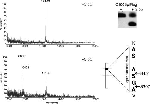
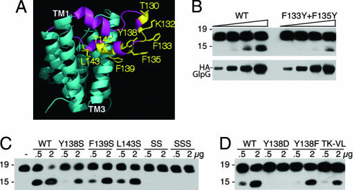
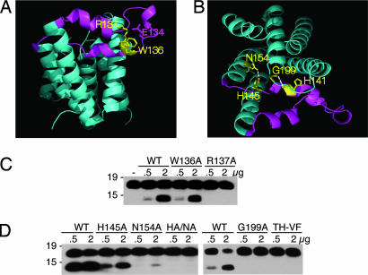
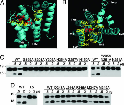
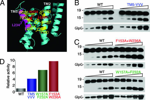
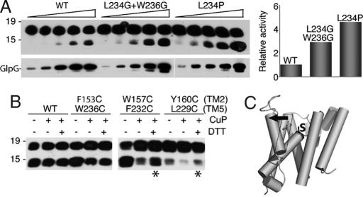
Intramembrane proteolysis is a core regulatory mechanism of cells that raises a biochemical paradox of how hydrolysis of peptide bonds is accomplished within the normally hydrophobic environment of the membrane. Recent high-resolution crystal structures have revealed that rhomboid proteases contain a catalytic serine recessed into the plane of the membrane, within a hydrophilic cavity that opens to the extracellular face, but protected laterally from membrane lipids by a ring of transmembrane segments. This architecture poses questions about how substrates enter the internal active site laterally from membrane lipid. Because structures are static glimpses of a dynamic enzyme, we have taken a structure-function approach analyzing >40 engineered variants to identify the gating mechanism used by rhomboid proteases. Importantly, our analyses were conducted with a substrate that we show is cleaved at two intramembrane sites within the previously defined Spitz substrate motif. Engineered mutants in the L1 loop and active-site region of the GlpG rhomboid protease suggest an important structural, rather than dynamic, gating function for the L1 loop that was first proposed to be the substrate gate. Conversely, three classes of mutations that promote transmembrane helix 5 displacement away from the protease core dramatically enhanced enzyme activity 4- to 10-fold. Our functional analyses have identified transmembrane helix 5 movement to gate lateral substrate entry as a rate-limiting step in intramembrane proteolysis. Moreover, our mutagenesis also underscores the importance of other residue interactions within the enzyme that warrant further scrutiny.


Conflict of interest statement

The authors declare no conflict of interest.

Figures

Fig. 1.
Fig. 1.
C100Spitz-Flag substrate is cleaved at two intramembrane sites. Pure C100Spitz-Flag was incubated for 2 h at 37°C with GlpG or buffer alone, and the C-terminal cleavage products were captured and analyzed in parallel by MALDI-TOF mass spectrometry (Left) and Western blot (Upper Right). The predicted mass of intact C100Spitz-Flag is 12,166 Da. The Spitz substrate motif (bracketed) is shown, with an external lysine N-terminal to the transmembrane domain above. Predicted masses of cleavage products designated by arrows are shown, and they correspond well to the peaks that appear in the mass spectrum after incubation with GlpG but not buffer alone.
Fig. 2.
Fig. 2.
L1 loop residues that face toward membrane lipids are required for protease activity. (A) Lateral view of GlpG (2NRF) with L1 loop in magenta and its residue side chains that are expected to contact membrane lipid highlighted in yellow. (B) Effect of changing F133 and F135 to tyrosine on protease activity was compared with wild-type GlpG in a limiting enzyme dilution series (amounts used for each were 100 ng, 200 ng, 400 ng, and 800 ng). Anti-Flag Western blot analyses are shown, with mass standards in kilodaltons depicted to the left of each panel. GlpG levels in the reactions were compared directly by anti-HA Western blot analysis. (C) Y138, F139, and L143 were mutated to serine, and the effect on protease activity against C100Spitz-Flag was assessed. SS is a double Y138S+F139S mutant, whereas SSS carries all three residues mutated to serine. Two different amounts of enzyme (in micrograms) were assayed for each GlpG variant for 1 h, with the highest level of GlpG being approximately equimolar to substrate levels, and resulted in almost complete substrate cleavage by wild type (to assess whether mutants abolished activity). (D) Mutation of Y138 to aspartate or phenylalanine, as well as a double mutant T130V+K132L (TK-LV), reduced activity.
Fig. 3.
Fig. 3.
Disrupting L1 loop:core interactions reduces protease activity. (A) Side view of GlpG (2NRF) with the L1 loop highlighted in magenta and the conserved WR motif within the L1 loop shown in yellow. R137 makes a series of hydrogen bonds to upper regions of the loop and to E134. (B) Top view of GlpG (2NRF) with the L1 loop:core hydrogen bonds between N154 of transmembrane domain 2 and H145 of the L1 loop, and the backbone of H141 of the loop and G199 above helix 4. (C) Mutagenesis of W136 had a mild effect on protease activity of GlpG, whereas changing R137 to alanine abolished protease activity. Western blot analysis of C100Spitz-Flag cleavage is shown. (D) The effect of disrupting L1 loop:core contacts by mutagenesis of H145 and N154 individually to alanine, or their double mutant (HA/NA), as well as G199 to alanine and H141 to phenylalanine with the neighboring T140 to valine (TH-VF), tested for proteolytic activity.
Fig. 4.
Fig. 4.
Importance of residue interactions neighboring the active site on protease activity. (A) Lateral view of GlpG (2NRF) with catalytic residues in red and neighboring transmembrane residues chosen for mutagenesis in yellow. (B) Top view of GlpG (2IC8) with catalytic residues in red and Loop5 (L5) residues chosen for mutagenesis in yellow. (C) Mutagenesis of residues G199 and Y205 to alanine and G257 to valine that are near the active site, and the catalytic S201 and H254 to alanine, abolished protease activity. Note that, under longer incubation, Y205A displayed residual proteolytic activity. Mutating N251, which interacts with Y205 in the closed form, to alanine reduced but did not abolish activity under prolonged incubation (data not shown) unless it was combined with Y205. (D) Mutating residues 243–250 of the L5 loop all to glycine (L5) resulted in a strong decrease in activity, whereas mutating L5 residues to alanine individually had mild effects.
Fig. 5.
Fig. 5.
Mutations in transmembrane helix 5 dramatically stimulate protease activity. (A) Lateral view of the open conformation of GlpG (2NRF) with catalytic serine and histidine in red, helix 5 in magenta, and helix 5 and helix 2 residues that form the interface highlighted in yellow. The L234 side chain that lies on the opposite face of helix 5 is shown in magenta. (B) Mutation of helix 5 residues L229, F232, and W236 to valine in a triple mutant increase protease activity ≈4-fold in an enzyme dilution assay (titrated 50–800 ng in 2-fold increments, also in C). GlpG levels in the reactions were assessed by anti-HA Western blot analysis (Lower). Note that fold stimulation was evident by both quantifying cleavage products for the same concentration of GlpG and comparing which dilution of the mutant yielded activity similar to wild type. (C) Mutation of helix 5 residue W236 and helix 2 residue F153 both to alanine increased enzyme activity ≈10-fold compared with wild-type GlpG, whereas alanine substitutions at 232 and 157 increased activity ≈7-fold. (D) Enzyme activity of wild type and GlpG mutants was quantified by densitometry for several different enzyme concentrations, and average fold increase was plotted.
Fig. 6.
Fig. 6.
Effect of increasing or decreasing transmembrane helix 5 flexibility. (A) Mutating L234, which is on the opposite face of helix 5 and is not involved in forming the helix 5:helix 2 interaction (see Fig. 5A), to proline increased enzymatic activity 5-fold, whereas changing both L234 and W236 to glycine increased activity ≈3-fold, compared with wild-type GlpG. GlpG levels were varied from 50 to 800 ng in 2-fold increments, and resulting enzyme amounts in the reaction were verified by anti-HA Western blot analysis. Activity stimulation was quantified by densitometry and is depicted graphically (Right). (B) Corresponding pairs of residues on helices 2 and 5 were changed to cysteine, and activity of the untreated, oxidized (CuP), and oxidized and subsequently reduced (DTT) enzymes were assessed. Asterisks denote enzyme activity that could be restored by DTT after oxidation. Note that the double mutant containing mutated Y160 had a lower enzymatic activity, as observed for this mutation in conjunction with other helix 5 mutants. (C) Model of intramembrane substrate gating by rhomboid enzymes: lateral tilting (black arrow) of the top of transmembrane helix 5 opens a path for substrate entry to the catalytic serine (labeled S).

Comment in

  • From rhomboid function to structure and back again.
    Lieberman RL, Wolfe MS. Lieberman RL, et al. Proc Natl Acad Sci U S A. 2007 May 15;104(20):8199-200. doi: 10.1073/pnas.0702745104. Epub 2007 May 9. Proc Natl Acad Sci U S A. 2007. PMID: 17494772 Free PMC article. No abstract available.

Similar articles

Cited by

References

    1. Brown MS, Ye J, Rawson RB, Goldstein JL. Cell. 2000;100:391–398. - PubMed
    1. Wolfe MS, Kopan R. Science. 2004;305:1119–1123. - PubMed
    1. Urban S. Genes Dev. 2006;20:3054–3068. - PubMed
    1. Li YM, Lai MT, Xu M, Huang Q, DiMuzio-Mower J, Sardana MK, Shi XP, Yin KC, Shafer JA, Gardell SJ. Proc Natl Acad Sci USA. 2000;97:6138–6143. - PMC - PubMed
    1. Takasugi N, Tomita T, Hayashi I, Tsuruoka M, Niimura M, Takahashi Y, Thinakaran G, Iwatsubo T. Nature. 2003;422:438–441. - PubMed

Publication types

MeSH terms