. 2007 May 8;104(19):8053-8.
doi: 10.1073/pnas.0611669104. Epub 2007 Apr 26.

GATA6 is an astrocytoma tumor suppressor gene identified by gene trapping of mouse glioma model

Affiliations

GATA6 is an astrocytoma tumor suppressor gene identified by gene trapping of mouse glioma model

Deepak Kamnasaran et al. Proc Natl Acad Sci U S A. .

Abstract

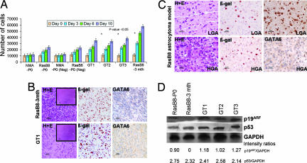
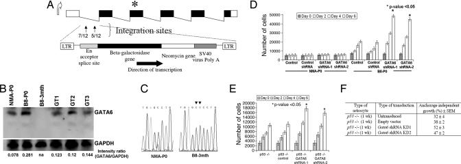
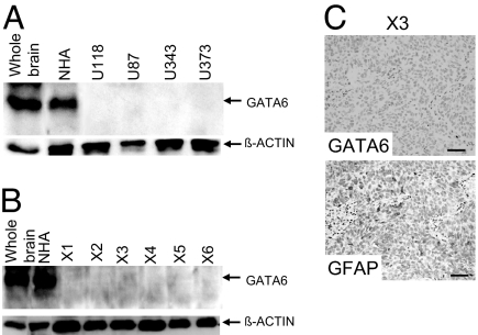
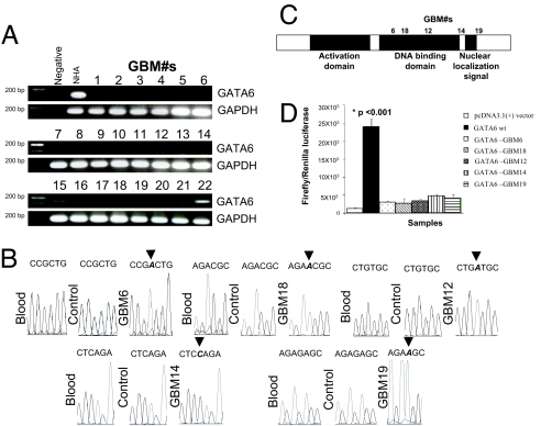
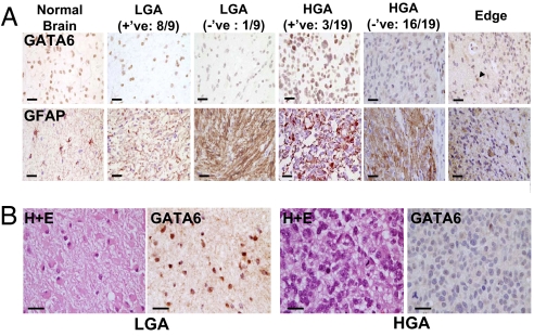
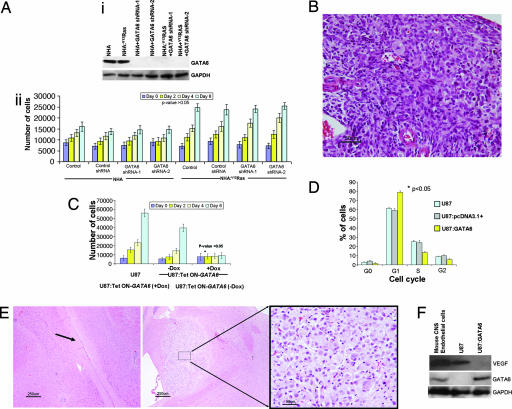
Malignant astrocytomas are the most common and lethal adult primary brain tumor. Retroviral gene trapping of nontransformed neonatal astrocytes from a glial fibrillary acidic protein (GFAP):(V12)Ha-Ras murine astrocytoma model led to isolation of the transcription factor Gata6. Loss of Gata6 resulted in enhanced proliferation and transformation of astrocytes. Human malignant astrocytoma cell lines, explant xenografts, and operative specimens demonstrated loss of GATA6 expression. Loss-of-function GATA6 mutations with loss of heterozygosity of the GATA6 locus were found in human malignant astrocytoma specimens but not in lower-grade astrocytomas or normal adult astrocytes. Knockdown of Gata6 expression in (V12)Ha-Ras or p53-/- astrocytes, but not in parental murine or human astrocytes, led to acceleration of tumorgenesis. Knockin GATA6 expression in human malignant astrocytoma cells reduced their tumorgenic growth with decreased VEGF expression. Collectively, these data demonstrate that GATA6, isolated from a murine astrocytoma model, is a novel tumor suppressor gene that is a direct target of mutations during malignant progression of murine and human astrocytomas. This work also demonstrates the utility of random mutagenesis strategies, such as gene trapping, on murine cancer models toward discovery of novel genetic alterations in corresponding human cancers.


Conflict of interest statement

The authors declare no conflict of interest.

Figures

Fig. 1.
Fig. 1.
Proliferation and tumorigenic properties of gene-trapped astrocytes derived from the B8 GEM astrocytoma model. (A) MTT proliferation assays of NMAs from CD1-ICR newborn pups (NMA-P0), newborn B8 pups (B8-P0), both transduced with empty vector controls (Neg), gene-trapped B8-P0 astrocyte clones (GT1, GT2, and GT3), and B8–3mth astrocytoma cultures derived from a mouse harboring a HGA. Gene-trapped B8-P0 clones have a proliferation advantage compared with parental and Neg NMA-P0 and B8-P0 astrocytes, approaching the proliferation rate of the transformed B8–3mth astrocytoma cultures. (B Upper) Pathological features of invasive malignant astrocytoma in the frontal cortex of Nod-Scid mice 1 month after injection of 106 B8–3mth astrocytes (H&E). β-Gal staining confirms expression of the GFAP:V12HaRas-IRES-LacZ transgene. GATA6 expression is lost in the HGA. (Lower) Pathological features of invasive malignant astrocytoma in the frontal cortex of Nod-Scid mice that developed in ≈7% of B8-P0 gene-trapped astrocytes (GT1 clone shown; H&E), with β-gal expression from both the GFAP:V12HaRas-IRES-LacZ transgene and the gene-trap vector. Parental or empty gene-trap vector-negative NMA and B8-P0 controls did not grow in Nod-Scid mice up to 6 months of observation. The finding of loss of Gata6 expression in the HGA suggests that an acquired mutation occurred in the nontargeted gene-trapped allele. (C) B8 GEM astrocytomas at the age of 1 month (LGA, Upper) and 3 months (HGA, Lower) (H&E). Gata6 is expressed in 1-month LGA (Upper Right) but absent in 3-month HGA (Lower Right), suggesting a role in tumor progression. β-Gal staining (Center) confirms expression of the GFAP:V12HaRas-IRES-LacZ transgene in the transformed astrocytes. GATA6 is abundantly expressed in wild-type age-matched brains (13). (Magnifications: ×400 for B and C and ×40 for Insets in B Left. Scale bars: 25 μm for B and C and 250 μm for Insets in B Left.) (D) Western blot analysis demonstrating that p19ARF and p53 expression is relatively unchanged in the B8 gene-trapped clones (GT1, GT2, and GT3) when compared with the B8-P0 parental cells. p19ARF expression is absent in the B8–3mth astrocytoma cells. Approximately 40 μg of protein lysates was loaded in each well. GAPDH was used to assess the amount of protein loaded.
Fig. 2.
Fig. 2.
Characteristics of gene-trapped astrocytes. (A) Schematic of the murine Gata6 transcript isoform expressed in the CNS. Integrations of the retroviral gene-trap vector were identified within the first intron of Gata6 and in the same orientation as the endogenous promoter. The number of integrations per site is shown in the first intron. ∗ represents the map position of a homozygous frameshift mutation identified in exon 3 of B8–3mth astrocytes. (B) Western blot analyses on protein lysates isolated from primary astrocyte cultures, with 40 μg of total protein lysate loaded per lane. Absent Gata6 expression is noted in B8–3mth astrocytoma cells (consistent with findings in Fig. 1 B and C). GAPDH was used as a positive control for loading and normalization of the densitometric analyses with Fluor Chem software. Gata6 expression was reduced between ≈50% and 58% among the three B8-P0 gene-trapped clones analyzed in detail (GT1, GT2, and GT3). (C) Chromatograms demonstrating a 1641_1642InsCC mutation in exon 3 of Gata6 in the B8–3mth astrocytes encoding the DNA binding domain. (D) Gata6 expression was constitutively knocked-down in NMA and B8-P0 astrocytes by ≈90% compared with controls by Western blot analysis (data not shown), with two shRNAs (mapping to murine exons 2 and 4) expressed from a stably integrated pSIREN-RetroQ vector. Parental or NMA and B8-P0 astrocytes engineered with a negative shRNA vector (control) were used as negative controls for the MTT proliferation assays. Gata6 knockdown induced a significant (∗, P < 0.05) proliferation advantage after day 2, only in the B8-P0 astrocytes and not in NMA. (E) Gata6 knockdown in homozygous null murine astrocytes induced a proliferative advantage (P < 0.05) and increased anchorage-independent growth in soft agar (F) compared with parental p53−/− astrocytes or those transduced with a negative shRNA vector (control).
Fig. 3.
Fig. 3.
Alterations in GATA6 expression among human GBM lines and explant xenografts. (A) Western blot analysis demonstrating loss of GATA6 expression in all four established human GBM lines. Each lane has 40 μg of total protein lysate, with β-actin used as a positive loading control. Whole-brain sample was obtained from a head injury patient requiring surgery, whereas NHA represents human hTERT immortalized but nontransformed astrocytes. (B) GATA6 expression was absent in six human GBM explant xenografts. Each lane has 40 μg of total protein lysate, with β-actin used as a positive loading control. (C) GATA6 expression in human GBM explant xenograft (X3) determined by IHC. A rabbit GATA6 polyclonal primary antibody and a protein G-HRP secondary antibody were used. None of the xenografts expressed GATA6, with 50% immunopositive for the differentiated astrocyte marker GFAP (rabbit GFAP polyclonal antibody from DAKO). No immunoreactivity was detected on the GBM explant xenograft specimens using only a secondary antibody. (Magnification: ×200.)
Fig. 4.
Fig. 4.
Screening of human GBM operative specimens for alterations in expression and mutations. (A) RT-PCR screening of a panel of 22 human GBM operative specimens. GATA6 expression was absent in 20/22 samples tested. GAPDH was used as a positive control marker. (B) Chromatograms of homozygous frameshift mutations identified in the GATA6 DNA binding domain and C terminus of five GBM specimens. These specimens also had loss of GATA6 RNA expression by RT-PCR (A), with loss of GATA6 protein expression in 19 specimens available for IHC evaluation (Fig. 5A). (C) Schemata of the human GATA6 protein with locations of homozygous frameshift mutations identified in the DNA binding domain (3/5) and C terminus (2/5) of GBM specimens and associated with LOH (SI Fig. 10). (D) DNA binding assays of GATA6 patient GBM mutations using the p450-Cytochrome:C17 promoter:pGL3 firefly luciferase reporter in U87 cells. Negative controls included nontransfected U87 plus transfection with the pcDNA3.1+ vector lacking only the GATA6 gene. The data (expressed as mean ± SEM) demonstrate that wild-type GATA6 induced significant expression (P < 0.001) of the firefly luciferase reporter from the p450-Cytochrome:C17 promoter. However, the GATA6 frameshift mutations identified from the GBM specimens resulted in minimal nonsignificant induction of the firefly luciferase reporter from the p450-Cytochrome:C17 promoter.
Fig. 5.
Fig. 5.
Screening of human astrocytomas operative specimens for alterations in expression using IHC. (A) GATA6 expression was tested by IHC in normal human brain (from head injury patient), nine LGA, and 19 human GBM paraffin-embedded specimens. One of nine (≈10%) LGA and 16/19 (≈85%) GBM had loss of GATA6 expression, suggesting that loss of GATA6 is involved in astrocytoma progression. Pictures show GATA6 (Upper) and GFAP (Lower) immunostaining. The rightmost panels demonstrate the invading edge of a GBM with loss of GATA6 expression, while nontransformed astrocytes and other cells retain GATA6 expression (arrowhead). (Magnification: ×400. Scale bars: 25 μm.) (B) GATA6 expression is analyzed in a patient with a pathologically documented secondary GBM. GATA6 expression is abundant in the initial resected LGA (Left) but absent when the patient recurred with a GBM (Right). Shown are H&E immunostaining and GATA6 immunostaining. (Magnification: ×400. Scale bars: 25 μm.)
Fig. 6.
Fig. 6.
Functional studies of human GATA6 shRNA knockdown and reexpression. (Ai) GATA6 expression was knocked down by two shRNAs (human exons 4 and 5) from a stably integrated pSIREN-RetroQ vector. Parental astrocytes and those expressing control shRNA were used as negative controls. (Aii) MTT proliferation assay on hTERT immortalized nontransformed NHA and NHA:V12Ha-Ras human astrocytes. GATA6 knockdown did not induce a significant in vitro proliferation advantage in the NHA or NHA:V12Ha-Ras cells (P > 0.05). (B) Intracranial injection into Nod-Scid mice with 106 NHA:V12Ha-Ras + GATA6 shRNA knocked-down astrocytes, but not controls, resulted in in vivo malignant astrocytomas (H&E; Magnification: ×200), demonstrating tumor cells with nuclear atypia and mitosis. (C) GATA6 reexpression induced antiproliferative effects on U87:Tet ON-GATA6 lines treated with Dox (P < 0.05). Leaky expression of GATA6 in the pREV-TRE vector can account for slight inhibition of proliferation even in untreated cells compared with parental U87 cells over the 6-day analysis period. (D) Cell cycle flow cytometry analysis of U87 cells transiently expressing GATA6 under control of the cytomegalovirus promoter (pcDNA3.1+). Nontransfected and empty vector (pcDNA3.1+) transient transfected U87 cells served as negative controls. Transient reexpression of GATA6 induced a significant (P < 0.05) increase in G1 arrested cells compared with the negative controls. (E) Intracranial xenografts with 106 U87:Tet ON-GATA6 cells with or without Dox in the drinking water. Tumor growth was not seen after ≈4 months in mice treated with Dox (Left; arrow shows injection tract). In contrast, mice without Dox had to be killed at 4 weeks, with development of large tumor nodules at the site of injection (Center; Right shows a magnified view of the tumor). (Magnification: ×40 in Left and Center and ×400 in Right. Scale bars: 250 μm in Left and Center and 25 μm in Right.) (F) GATA6 regulation of VEGF expression. U87 cells express VEGF (similar to control mouse brain endothelial cells) but not GATA6. Transient reexpression of GATA6 in the U87 cells inhibits VEGF expression.

Comment in

  • Trapping the mouse genome to hunt human alterations.
    Bleau AM, Holland EC. Bleau AM, et al. Proc Natl Acad Sci U S A. 2007 May 8;104(19):7737-8. doi: 10.1073/pnas.0702617104. Epub 2007 May 3. Proc Natl Acad Sci U S A. 2007. PMID: 17483476 Free PMC article. No abstract available.

Similar articles

Cited by

References

    1. Mahaley MS, Jr, Mettlin C, Natarajan N, Laws ER, Jr, Peace BB. J Neurosurg. 1989;71:826–836. - PubMed
    1. Kleihues P, Cavenee WK, editors. WHO Classification of Tumors: Pathology and Genetics of Tumours of the Nervous System. Lyon, France: Int Agency for Res on Cancer; 2000.
    1. Stupp R, Mason WP, van den Bent MJ, Weller M, Fisher B, Taphoorn MJ, Belanger K, Brandes AA, Marosi C, Bogdahn U, et al. N Engl J Med. 2005;352:987–996. - PubMed
    1. Louis DN, Gusella JF. Trends Genet. 1995;11:412–415. - PubMed
    1. Guha A, Feldkamp MM, Lau N, Boss G, Pawson A. Oncogene. 1997;15:2755–2765. - PubMed

Publication types