. 2007 Sep;37(3):264-72.
doi: 10.1165/rcmb.2006-0418OC. Epub 2007 Apr 26.

Airway hyperresponsiveness, remodeling, and smooth muscle mass: right answer, wrong reason?

Affiliations

Airway hyperresponsiveness, remodeling, and smooth muscle mass: right answer, wrong reason?

Madavi N Oliver et al. Am J Respir Cell Mol Biol. 2007 Sep.

Abstract

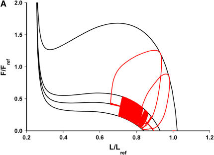
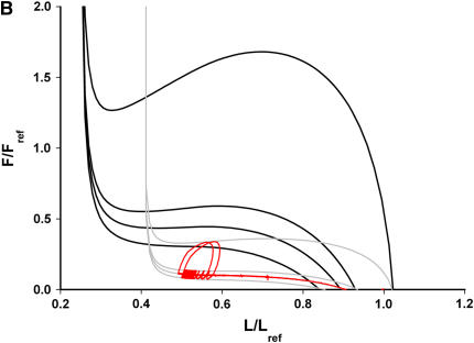
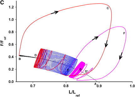
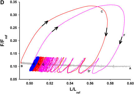
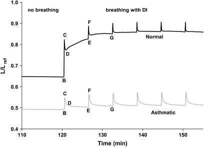
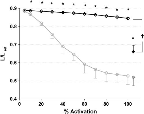
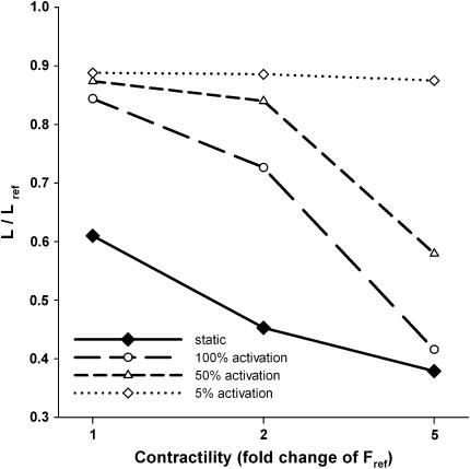
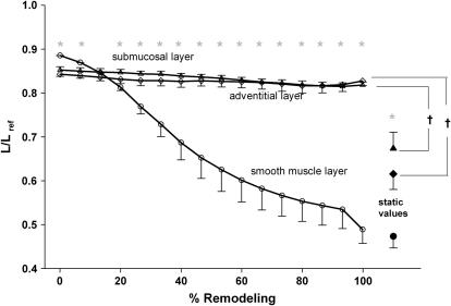
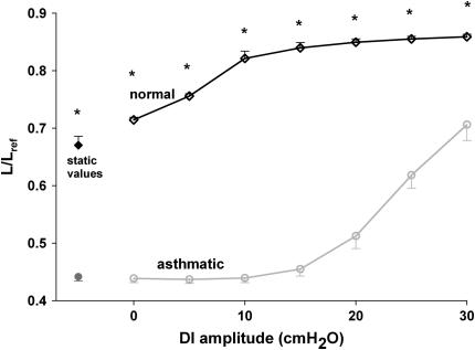
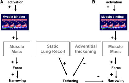
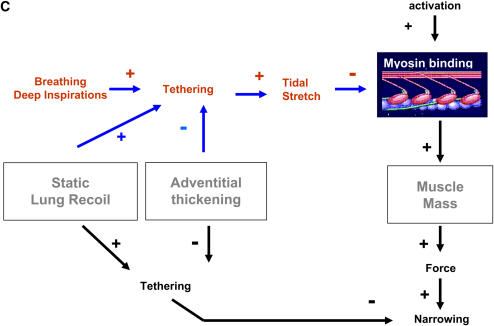
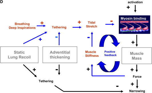
We quantified the effects of airway wall remodeling upon airway smooth muscle (ASM) shortening. Isolated ASM from sheep was attached to a servo-controller that applied a physiologic load. This load could be altered to reflect specified changes of airway wall geometry, elasticity, parenchymal tethering, transpulmonary pressure (P(L)), and fluctuations in P(L) associated with breathing. Starting at a reference length (L(ref)), ASM was stimulated with acetlycholine and held at constant P(L) of 4 cm H(2)O for 2 h. When all compartments were thickened to simulate the asthmatic airway but P(L) was held fixed, ASM shortened much more than that in the normal airway (to 0.52 L(ref) versus 0.66 L(ref)). When breathing with deep inspirations (DIs) was initiated, within the first three DIs the ASM in the normal airway lengthened to 0.84 L(ref), whereas that in the asthmatic airway remained stuck at 0.53 L(ref). Thickening of the smooth muscle layer alone produced the greatest muscle shortening (to 0.47 L(ref)) when compared with thickening of only submucosal (to 0.67 L(ref)) or only adventitial (to 0.62 L(ref)) compartments. With increased ASM mass, the ASM failed to lengthen in response to DIs, whereas in the airway with thickened submucosal and adventitial layers ASM lengthened dramatically (to 0.83 L(ref)). These findings confirm the long-held conclusion that increased muscle mass is the functionally dominant derangement, but mechanisms accounting for this conclusion differ dramatically from those previously presumed. Furthermore, increased ASM mass explained both hyperresponsiveness and the failure of a DI to relax the asthmatic airway.


Figures

<b>Figure 1.</b>
Figure 1.
Load characteristics for different transpulmonary pressures (PL) from normal muscle (dark lines) and asthmatic muscle (light lines). Smooth muscle length L was plotted as a fraction of Lo (radius of relaxed smooth muscle at a PL of 10 cm H2O) and active force F was plotted as a fraction of Fo (maximum force the muscle can generate, computed using the thickness of the smooth muscle at Lo and an estimated maximal stress of 150 kPa [12]). If the muscle generates a very high force, the airways are almost completely closed. If the muscle generates no force, the airways are nearly open. In between these extremes, the muscle force and length depend on pleural pressure, that is, the force and length of the muscle must lie on this line. Unlike the normal airway, the asthmatic airway already closes at a muscle length of 0.4 Lo, and muscle shortening requires much less force.
<b>Figure 2.</b>
Figure 2.
Force–length measurements from tracheal smooth muscle strips (red lines) and corresponding load characteristics (light and dark lines) for normal (A, C) and asthmatic airways (B, D). Activated muscle strips were statically equilibrated for 2 h at PL of 4 cm H2O, after which tidal breathing was imposed (sinusoidal fluctuations of amplitude 1.25 cm H2O with a single breath of amplitude 10 cm H2O every 6 min). A was the starting point, where the muscle was completely relaxed. After 2 h of static loading, the muscle shortened from A to B. BCD was the response to the first DI, and continued breathing opened the airways from D to E. EFG was the response to the second DI, and the airway continued to relax. Overall, the muscle was driven toward progressively greater lengths and smaller forces. In contrast, asthmatic airways did not lengthen between DIs (E < D). The airways appeared to be stuck at static levels, and the smooth muscle experienced smaller force–length excursions.
<b>Figure 3.</b>
Figure 3.
Muscle length (averaged over the duration of one breath) versus time. When breathing and deep inspiration started, the normal airway (dark line) dilated in response to a DI and remained dilated, whereas the asthmatic airway (light line) only dilated by a small amount and quickly returned to its static state.
<b>Figure 4.</b>
Figure 4.
Muscle length averaged over one breath versus %activation in normal and asthmatic airways (n = 5 for each type of airway). After 30 min of full activation, the activation level was gradually reduced. This was simulated by equating the full force produced by the muscle to less and less tensile stress. While the normal airway is almost fully dilated after the first two DIs (significant when compared with the corresponding static values, P < 0.01), the asthmatic airway showed very little reaction. Only when the activation level was decreased below 15% did the asthmatic muscle lengthen to a level that was observed in normal airways at 100% activation. Error bars denote SE between muscle strips and the asterisk indicates a significant difference between the normal and asthmatic conditions (P < 0.05) at each level of activation.
<b>Figure 5.</b>
Figure 5.
Muscle length averaged over one breath versus contractility in normal airways (n = 3). Three different levels of force generation were used. The muscle was loaded statically for 2 h, after which breathing with DI was turned on. After 30 min of breathing, the activation level was gradually reduced. In response to static loading, the smooth muscle with the greatest force-generating capacity shortened the most. In response to breathing and DI, the muscle with increased force-generating capacity (5-fold) behaved like an asthmatic muscle and remained short till it was deactivated below 15%.
<b>Figure 6.</b>
Figure 6.
Muscle length averaged over one breath versus remodeling (n = 4 for each layer). After 2 h of static loading and 30 min of breathing, each layer was “unremodelled” individually. In response to static loading, the airway with asthmatic smooth muscle layer shortened the most (P < 0.01). The smooth muscle layer played the greatest role in reproducing hyperresponsiveness when compared with adventitial and submucosal layers. Error bars denote SE between muscle strips and the asterisk indicates a significant difference (P < 0.01) between the three different layers. The dagger indicates a significant difference (P < 0.01) between the static equilibrium values and the response to 30 min of breathing and DI for the adventitial and submucosal layers.
<b>Figure 7.</b>
Figure 7.
Muscle length averaged over one breath versus DI amplitude in normal and asthmatic airways (n = 4 for each case). After 2 h of static loading at PL = 4 cm H2O, tidal breathing (sinusoidal fluctuations of amplitude 1.25 cm H2O) was imposed. There was no DI for the first 30 min. Thereafter a DI was introduced every 6 min. The DI amplitude was varied in steps of 5 cm H2O, from 5–30 cm H2O, every 30 min. In response to breathing the muscle in the normal airway lengthened immediately (even in the absence of DI) and it continued lengthening by a small amounts with increasing DI amplitudes. The muscle in the asthmatic airway remained at static lengths and only lengthened in response to a DI amplitude of 20 cm H2O and more. Error bars denote SE between muscle strips, and the asterisk indicates a significant difference between the normal and asthmatic conditions (P < 0.01) at each DI amplitude.
<b>Figure 8.</b>
Figure 8.
(A) The qualitative picture before 1992: the picture of airway narrowing was qualitative but predicted intuitively that greater muscle mass would lead to greater active force and, hence, greater airway narrowing. (B) Balance of static forces: Wiggs and colleagues (11) and Lambert and coworkers (12) performed the first quantitative analysis of airway narrowing. Their analysis rests upon a balance of static forces, with muscle length being set by the balance of active force generated by the muscle versus the passive elastic load against which the muscle is shortening. Although remodeling of various airway compartments were shown to decrease the load and thus contribute importantly to the extent of airway narrowing, the dominant effect was increased muscle mass. (C) Tidal loading: In the mid-1990s many investigators recognized that tidal loading is a potent inhibitor of active muscle forces. While this picture helped to explain why tidal breathing and deep inspirations are potent bronchodilators, it failed to explain why individuals with asthma are refractory to the beneficial effects of a DI. (D) Myosin dynamics: Due to a virtuous positive feedback loop, breathing is seen to be good for breathing. This positive feedback ensures that, even during maximal muscle stimulation, tidal stretches and/or DIs act to perturb the binding of myosin to actin, and thus keep active force and muscle stiffness quite small. Small muscle stiffness, in turn, keeps the muscle highly responsive to tidal loading, and so on. However, with decreased static lung recoil, increased adventitial thickening, or, especially, increased muscle mass, the myosin binding is perturbed less, the muscle becomes stiffer as a result, and stretches even less, and so on. The feedback loop collapses and the muscle becomes so stiff as to be refractory to the effects of DIs. “+” and “−,” respectively, indicate phenomena that act to increase or decrease the indicated effect. For example, adventitial thickening decreases tethering forces, and tethering forces act to decrease the extent airway narrowing. By decreasing tethering, therefore, adventitial thickening acts to exacerbate airway narrowing.

References

    1. Woolcock AJ, Peat JK. Epidemiology of bronchial hyperresponsiveness. Clin Rev Allergy 1989;7:245–256. - PubMed
    1. McParland BE, Macklem PT, Pare PD. Airway wall remodeling: friend or foe? J Appl Physiol 2003;95:426–434. - PubMed
    1. Macklem PT. Mechanical factors determining maximum bronchoconstriction. Eur Respir J Suppl 1989;6:516s–519s. - PubMed
    1. Sterk PJ, Bel EH. Bronchial hyperresponsiveness: the need for a distinction between hypersensitivity and excessive airway narrowing. Eur Respir J 1989;2:267–274. - PubMed
    1. Bai TR, Knight DA. Structural changes in the airways in asthma: observations and consequences. Clin Sci (Lond) 2005;108:463–477. - PubMed

Publication types